Seeing Cells on the Web
INTRODUCTION
Cells are the fundamental unit of life and disease; therefore, many avenues of research converge on cells, making images of cells prominent in research and teaching. Besides, cells are beautiful, and a gorgeous photo of a cell can spice up an otherwise dreary talk. Much of the progress of modern biomedical science can be tied to advances in our ability to better visualize the functional morphology of cells, including higher resolution imaging, informative molecular tags, and techniques for making observations in living cells. Given the microscopic scale that cells inhabit, and the variety of imaging techniques used to visualize them, it's not surprising that students are both inspired and confused by cells. How big are they? What shapes can they exhibit? How do cells perform their myriad functions? How do scientists produce all those pretty colors? This Feature describes websites that would help answer these and other important questions about cells, as well as sites that would help address some common sources of difficulty in the presentation of cellular information.
THE POWER OF 10
Let's begin with the issue of scale. A common problem for many students in understanding the sizes of cells is photomicrographs without scale bars, or unlabeled scale bars, or changing scale bars to make matters worse. On the other hand, scale bars can mar a beautiful image, and students often ignore the information they provide. Scale in the natural world is important and interesting enough to be worthy of explicit discussion with students. Many readers may be familiar with the classic film Powers of 10 by Charles and Ray Eames. I first saw the film when it came out in 1977 and I was in high school. The film made a lasting impression on me, in part because the setting is a park in my native Chicago. The film has been paid homage in many creative ways over the years, including a beautiful IMAX version and a hilarious animated feature of a child on a pogo stick jumping by powers of 10, over the hedge, over the rooftops, and then out into the universe. The official Eames Powers of 10 website (www.powersof10.com) makes the original film available, along with some other useful features (Figure 1). Sample images at various powers of 10 are provided, along with links to websites concerned with any given scale. You click on 10−5 or 10−6, for example, and see cellular images and can then follow links to other websites addressing the world at that scale. It is a wonderful idea, but unfortunately the links appear to be somewhat haphazardly chosen and some are out of service. To fully use the site, active X controls and the Shockwave plug-in are required.

Figure 1. The Powers of 10 website (www.powersof10.com) features the classic 1977 Eames film.
BEYOND THE BAG OF JELLO
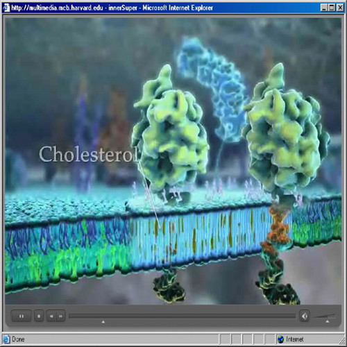
Cells are tiny, and yet they are nearly incomprehensibly complicated. To paraphrase Lewis Thomas in The Lives of a Cell, a cell isn't like a little chemical factory; a cell is like a crowded metropolis, packed with structures and conveyances frenetically shuttling goods. In an effort to simplify and teach clearly, various organelles are usually treated in isolation, the interactions between structures and molecules are reduced to the minimum number of players, and students can get the impression that a cell's activity is carefully orchestrated by invisible hands. Fortunately, a powerful animated antidote has recently become available through the Department of Molecular and Cellular Biology at Harvard. The “Inner Life of the Cell” and related animations (multimedia.mcb.harvard.edu/media.html) were produced by Alain Veil, Robert Lue, and colleagues for use in undergraduate courses (Figure 2). Animators like John Liebler of XVIVO and Drew Barry of WEHI TV and DNAi.org are innovators in a cinema verité style that evokes what things might really be like inside a busy cell. The “Inner Life of the Cell” is a five-minute tour de force that should engage the most jaded student and inspire dedicated biophiles. Although highly engaging animations can teach specific concepts and facts about cells, their main value is to engage and inspire and to emphasize the astounding complexity of cellular processes and structures. Photorealistic animations are not without drawbacks; for example, students might infer that an animation, which is easy to understand, is more accurate than micrographs or other first-order data.

Figure 2. The Inner Life of the Cell animation is cinema verité for cell biology (multimedia.mcb.harvard.edu/media.html).
Animations are powerful, but they are representations that synthesize experimental observations while making numerous assumptions and simplifications. Cell biologists and microscopists have been making real movies of living cells for decades, and there are some excellent sources of movies on the Web. The publisher of CBE—Life Sciences Education, the American Society of Cell Biology (ASCB), publishes a website called the ASCB Image & Video Library (http://cellimages.ascb.org/). This fledgling website has only 12 movies so far, but they are excellent, like the supremely beautiful “Dance of the Chromosomes.” Most of the movies are in the “Nucleus” section, which also has great still images. There are 40 resources in the “Cytoplasm” section, including some movies. The oddly named “Founders” category betrays the historical emphasis of the website, which is heavy on electron microscopic (EM) images. The rigorous documentation of each resource is welcome, and although the historical approach has limited appeal, it has obvious value, at least to instructors. The site has useful features designed to make the images easily adaptable for use in presentations and publications, like the ability to zoom and rotate images. And fortunately, scale bars are prominent and well labeled! The ASCB library is run like a true collection with a curator, peer reviewers, and accession notes. I think in the future, when this site has 100 videomicroscopy movies, 100 animations, and 1000 photos, the categories and indexing features will naturally improve. The video library is a potential treasure that should be supported, and they are looking for both reviewers and contributions.
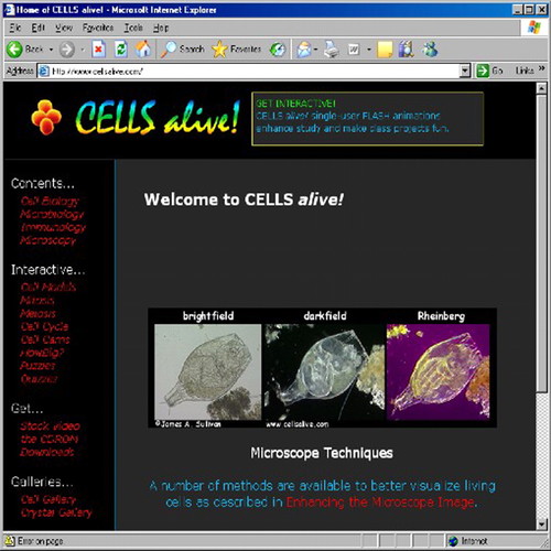
Movies are also available on the popular Cells Alive! website (www.cellsalive.com). Jim Sullivan's website has been winning kudos since the mid-1990s (Figure 3). Many good movies are available to freely view on the website, but many more movies are for purchase. The site has a snappy design and an excellent search function, and I found the jigsaw puzzles a welcome diversion. The animations on the site are highly schematic and run too fast, but they do have a frame-by-frame step-through feature. The mitosis animation has the smart concept of simultaneously showing a movie of a dividing cell along with an animation of cell division, but the movie is too small, and the animation too sketchy. On the other hand, the simple cell-cycle animation was effective and clear. There are tutorials on prokaryotic cells and on the parts of plant and animal cells that feature some simple mouse-over interactivity. Clicking on organelles or their names produces new text explanations that are good at reminding students of the techniques used to determine a particular structure and its appearance, for example, the fact that rough endoplasmic reticulum is named for its “pebbled” appearance in EM due to ribosomes on the surface.

Figure 3. Cells Alive! (www.cellsalive.com) has been a popular source of cell biology movies, photos, and tutorials since the early days of the Web.
ALL THE PRETTY COLORS
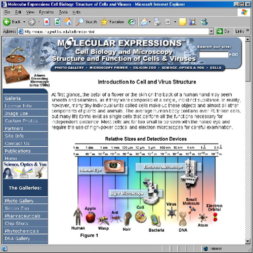
One of the best websites for finding detailed information on various optical methods, digital enhancements, and fluorescent reagents is Michael Davidson's Molecular Expressions (http://micro.magnet.fsu.edu) affiliated with Florida State University and the National High Magnetic Field lab (Figure 4). The site makes extensive use of JAVA for demonstrations and tutorials. The section on “Live-Cell Imaging Techniques,” for example, has detailed information on keeping cells alive during observation, special optical pathway and detector requirements, automated observation, and vital fluorescent probes. The movies in this section are very good as well. The coverage of imaging technology and techniques is comprehensive, from the physics of standard light microscopy to how to set up a near-field scanning optical microscope. To explain all those pretty colors we see, here's just a sample of the topics covered in the section on Fluorescence: Introductory Concepts, Fundamentals, Microscope Anatomy, Filters, Light Sources, Focusing and Alignment, Electronic Imaging Detectors, Fluorophores, Fluorescent Proteins, Indirect Immunofluorescence, Photomicrography, Multiphoton Excitation Microscopy, Fluorescent Resonance Energy Transfer, Total Internal Reflection Fluorescence Microscopy, Laser Systems, Stereomicroscopy, and many others. Although the site has fantastic resources, it is text-heavy and has some navigation and organization problems. For example, I could not find the nice powers of 10 tutorial (http://micro.magnet.fsu.edu/primer/java/scienceopticsu/powersof10/) from the homepage or from the site search, though I was successful with a Google search.

Figure 4. Molecular Expressions (http://micro.magnet.fsu.edu/) provides rigorous explanations of all the major methods for visualizing cells, as well as images, movies, and JAVA tutorials.
TIP OF THE ICEBERG OR ONION
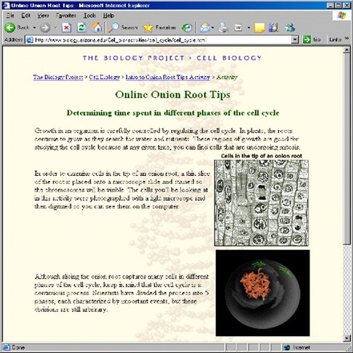
Beautiful images can be so captivating that we may forget that pictures of cells provide scientific information; images are data. As an undergraduate, I was dubious about the notion that I could learn anything about cellular functions by looking at fixed and sectioned cells. But scoring and counting cells at various stages of mitosis in a plant root tip to calculate the time a cell spent at various stages in mitosis taught me a lot about sampling, the power of simple calculations, and how to extract functional information from snapshot data. I was hoping to find a version of this classic experiment on the Web and my quest was rewarded by a resource at the University of Arizona. The University of Arizona Biology Project has an excellent set of cell biology tutorials (www.biology.arizona.edu/Cell_bio/cell_bio.html). The tutorials feature good graphics and some photos, covering membranes, signaling, cytoskeleton, cell cycle, cell division, and the intracellular organization of cells. The website has a very nice simulation of the classic cell-cycle calculation exercise. Following an optional tutorial on mitosis, I discovered a series of onion cell micrographs and was tasked to categorize each cell as being in interphase, prophase, metaphase, anaphase, or telophase (Figure 5). I did pretty well. The activity includes creative feedback for both correct and incorrect answers. After classifying more than 30 cells, I was asked to make the calculations to estimate how much time in each phase a cell spends. How well does this method work? How prone is it to sampling errors? Send your students to the Rutgers University website (http://bio.rutgers.edu/∼gb101/lab2_mitosis/highmagroot1.html) to see a gorgeous root tip section to calculate an independent sample for comparison. In answer to students who think the exercises are merely academic busy work, have them go to the University of Tennessee website (www.tiem.utk.edu/∼gross/bioed/webmodules/tumorgrowth.html) for information on estimating the growth rate of tumors as a biomedical analogous application. For an update on the state of the art for extracting useful quantitative data from cellular images, visit CellProfiler (http://www.cellprofiler.com/) to see sample experiments and images and to download free software.

Figure 5. The University of Arizona has a nice online version of the classic onion root tip mitosis lab exercise (http://www.biology.arizona.edu/Cell_bio/activities/cell_cycle/cell_cycle.html).
OTHER NOTEWORTHY WEBSITES
North Dakota State University's Virtual Cell animation collection (http://vcell.ndsu.nodak.edu/animations/) began as an online game aimed at undergraduates. The creators soon realized, however, that the beautiful 3D graphics that they had created for the game environment would also be useful in more standard didactic settings. The First Look, Advanced Look, and Run Animation options, are a simple and very smart idea that animation developers should consider adopting more widely. In the First Look and Advanced Look, snapshots from the animations are presented in storyboard manner to identify key players and explain briefly the action, with the Advanced Look doing so in more detail. My intuition that these viewing options should be an effective method to increase learning, retention, and understanding is supported by assessment data (McClean et al., 2005). Unfortunately, the animations are only available in Windows Media format, a drawback given the high number of Macintosh users in life sciences education.
The following websites from Emory University, University of Washington, Queen's University in Ontario, and the University of Delaware have good information and images if you are particularly interested in confocal microscopy: http://www.physics.emory.edu/∼weeks/confocal/; http://depts.washington.edu/keck/intro.htm; http://meds.queensu.ca/qcri/flow/cri-cm.htm; and http://www.udel.edu/Biology/Wags/wagart/confocalpage/confocal.html.
The Micropolitan Museum from the Institute for the Promotion of the Less Than One Millimeter (www.microscopy-uk.org.uk/micropolitan/index.html) is a clever virtual museum of microscopic creatures. Visit “the floorplan” to fully appreciate curator Wim van Egmond's concept.
Cells! and ER! (www.danforthcenter.org/Cells/) websites from the Danforth Plant Science Center produced by R. Howard Berg have a nice collection of plant cell images and some movies made from serial section reconstructions.
An online resource for cell and molecular biologists, Cell & Molecular Biology Online (www.cellbio.com/images.html), has a set of links to many online collections of images.
Micromacro is a nicely executed website by photographer Sinclair Stammers (www.micromacro.co.uk/index.htm). The site has beautiful images of living forms including cells and a simple, well-designed viewing console.
A student and teacher collaboration website (http://library.thinkquest.org/12413/) from the Oracle Education Foundation competition is a very nice example of student work featuring good drawings and links for cell biology.
As always, comments and suggestions for future topics are welcome and can be sent to Dennis Liu at dliu@hhmi.


