Realms of the Viruses Online
INTRODUCTION
What better time than the flu season to explore the realms of the viruses? From your nasal passages to the dirt under your feet, viruses are everywhere, in innumerable hordes. They display a dazzling array of geometries, from compact rods to voluminous dodecahedrons (12-sided polyhedron). While all viruses are small compared with our cells (∼10,000 nm in diameter), they range over many magnitudes, from the tiny poliovirus (30 nm in diameter) to the bacterial-sized poxvirus (450 nm in diameter). Viruses have evolved strategies for infecting all taxa, but most viruses are highly specific about their cellular host. In humans, viruses cause diverse diseases, from chronic but benign warts, to acute and deadly hemorrhagic fever. Viruses have entertaining names like Zucchini Yellow Mosaic, Semliki Forest, Coxsackie, and the original terminator, T4. But what I find most fascinating about viruses is their testimony to the power of information. To understand viruses, we need to explore and understand their structure, and we need to interrogate their genomes. In this article, I will review a number of websites that open a portal to the realms of the viruses.
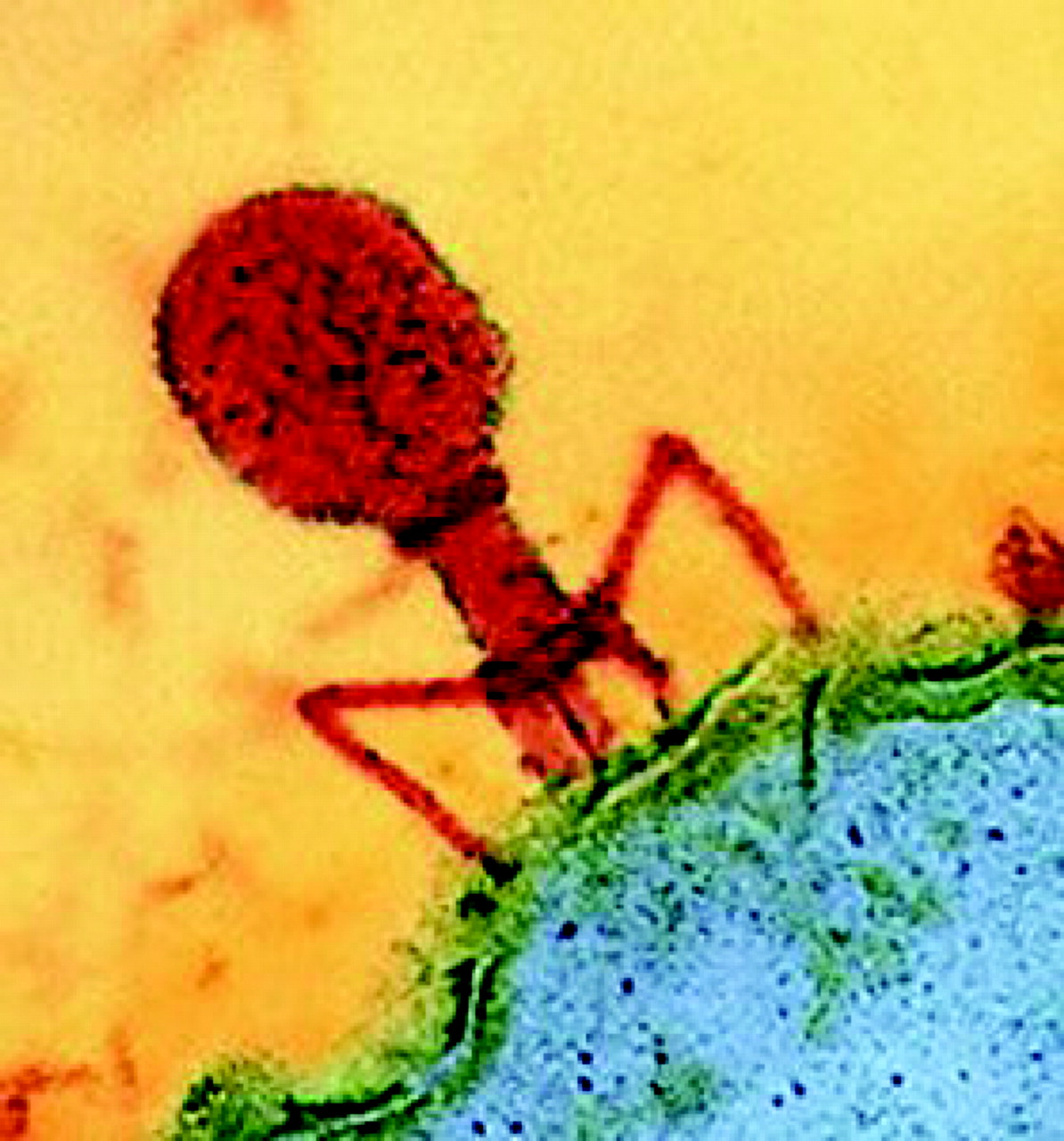
Figure 1. A colorized image of T4 bacteriophage at a cell's surface from Wikimedia (http://commons.wikimedia.org/wiki/Main_Page). The image accompanied an online article on the therapeutic potential of viruses (http://www.cosmosmagazine.com/node/1024) from Cosmos magazine.
Let's begin with a look at influenza. The U.S. Centers for Disease Control and Prevention (CDC) has some excellent information on “the flu” at www.cdc.gov/flu, including dispelling the myth of the “stomach flu,” a completely unrelated disease not of viral origin. The CDC website is huge and document heavy, so searching on keywords is often the best start. A search generally leads you to large, fairly well-organized sections on a given topic. The CDC's Key Facts page on the flu (http://www.cdc.gov/flu/keyfacts.htm) tells the reader that in an average season as much as 20% of the U.S. population succumbs to the flu, 200,000 Americans are admitted to a hospital, and 36,000 die. Data collected since 1982 show that the flu is clearly more prevalent during the winter, with peak infection rates in February most years, but December, January, and March have often been the worst months while November was the peak month only once in 24 seasons (http://www.cdc.gov/flu/about/disease.htm). The seriousness and recurring seasonality of the flu epidemic should get students' attention and pique their curiosity. Why does a flu epidemic occur every year? Why does the flu vaccine change every year, and why is its effectiveness variable? Why is there a vaccine for influenza and not for the common cold? There's no one website to find all the answers, but on the Howard Hughes Medical Institute's (HHMI) BioInteractive pages (www.hhmi.org/biointeractive/disease/click.html) students will find an animation called Viral Subunit Reassortment that provides an introduction. What the animation lacks in 3D pizzazz is made up for by Don Ganem's lucid narration of how viral diversity is generated and enables influenza to dodge the immune system and vaccines. The animation was prepared for an HHMI Holiday Lecture that Dr. Ganem delivered before an audience of high school students, and it is worth viewing extended sections of his discussion of influenza. Go to http://www.hhmi.org/biointeractive/disease/lectures.html, select lecture 4, and click on the chapters of interest in the webcast window. A good source for more information on the influenza virus, including some photos and graphics, is John Kimball's online biology textbook's influenza chapter (http://users.rcn.com/jkimball.ma.ultranet/BiologyPages/I/Influenza.html). To get closer to the cutting edge of what biotechnology is doing to fight the flu, students could visit The Institute for Genomics Research website, http://www.tigr.org/viralGenomics.shtml, to learn about their joint project with the National Institutes of Health to sequence the genomes of hundreds of flu virus variants.

Figure 2. Electron micrograph of influenza virus particles from the CDC website, http://www.cdc/gov/flu/about/disease.htm.
Lifestyles of the Viruses, Especially HIV
Students studying influenza should note that the flu virus has an RNA genome, which has ramifications for how the virus interacts with its cellular host to highjack cellular machinery for viral reproduction. How do viruses' various types of genomes replicate? Blackwell Publishing has a set of animations to accompany its Basic Virology textbook, and Web versions can be found at http://www.blackwellpublishing.com/wagner/animation.asp#. The animation entitled Genome runs in Flash with graphics that are a bit too schematic for my taste. However, it is very useful to see the genome replication strategies of six different viruses (herpes simplex virus, poxvirus, polio, influenza, reovirus, and HIV) with different types of genomes. The Flash console enables you to view the animations repeatedly and to switch easily from virus to virus for comparison. A student will quickly grasp the implications of the genomic diversity of viruses. Of course, a viral genome will not be replicated unless the virus can first attach to, and then penetrate, a cell. The Blackwell animation titled Attach compares four different penetration modes: nonenveloped virus translocation, membrane fusion by an enveloped virus, endocytosis of an enveloped virus, and vector-mediated viral penetration of a plant cell wall. Again, the most useful aspect of the highly schematic animation is the side-by-side comparison.
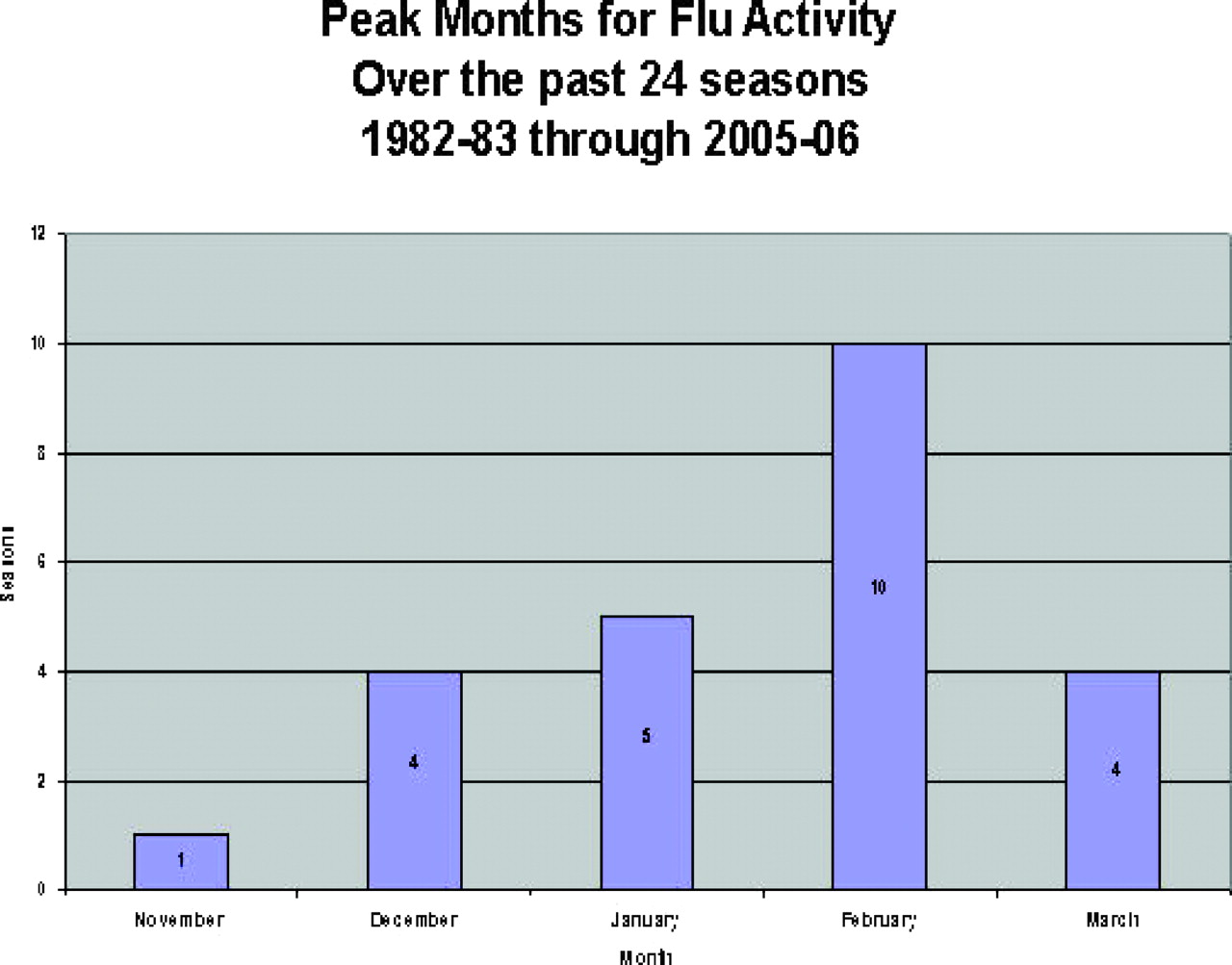
Figure 3. Data from the Influenza section of the CDC website, showing the peak months for influenza infections for 24 flu seasons.
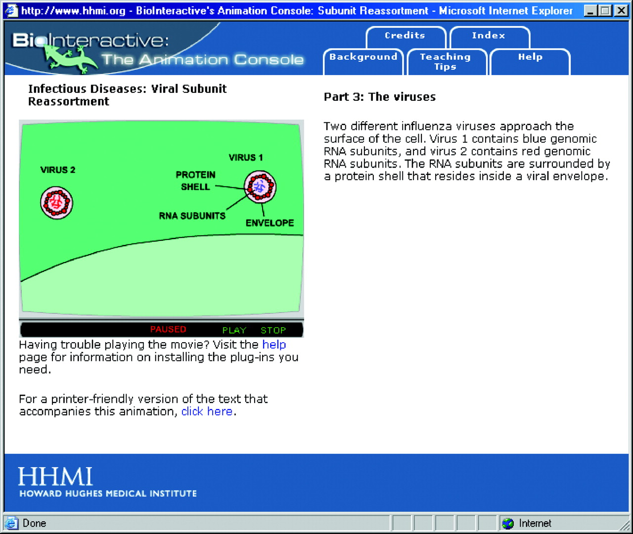
Figure 4. The Viral Subunit Reassortment animation on HHMI's BioInteractive shows how the RNA subunits of the influenza virus can re-assort to produce viral diversity that evades vaccines and the immune system.
Students may notice that while influenza and HIV both have RNA genomes, the way these genomes are replicated are entirely different, indicating divergent evolution and hosts. The life cycle of a retrovirus like HIV is more complicated than that of an RNA virus that doesn't integrate into the host genome. Influenza generates diversity by recombining genome segments in the cytoplasm, whereas HIV generates diversity via an error-prone reverse transcription step. The ensuing mutations of HIV present a moving target for medicines, as well as for vaccines and the host's immune system. Furthermore, retroviruses find safe haven by integrating into the host cell's genome, making eradication of the virus nearly impossible. Given the scope and importance of the worldwide AIDS epidemic, it is useful for students to learn more about the HIV life cycle and to appreciate how understanding the fundamentals of viral life cycle is the key to designing effective drugs and vaccines.
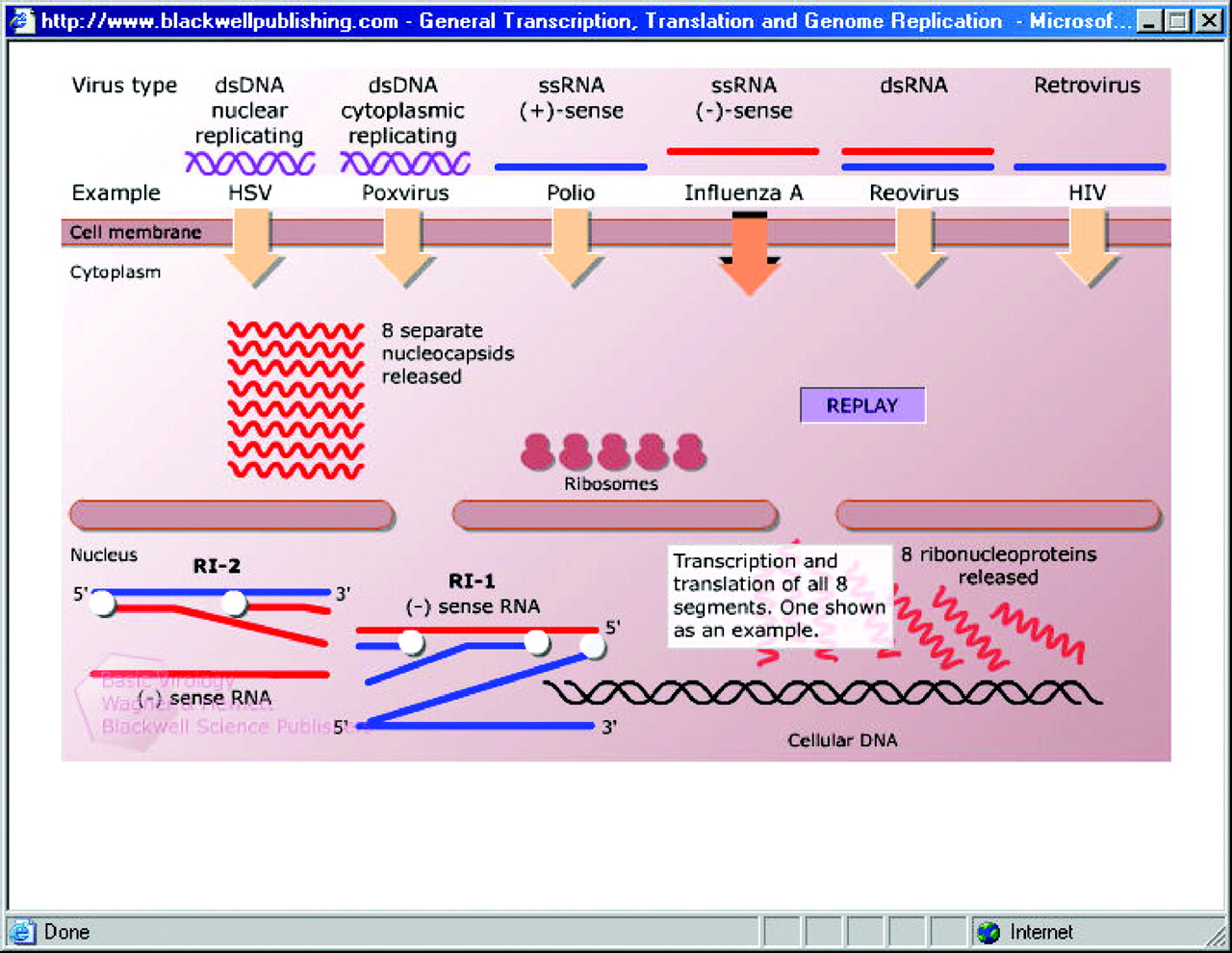
Figure 5. An animation from Blackwell Publishing compares the replication strategies of six different viruses.
An excellent 6-min animation of the HIV life cycle, emphasizing drug targets, is available on the National Academy of Sciences Koshland Science Museum website at http://www.koshland-science-museum.org/exhib_infectious/hiv_antivirals_movie1.jsp. The animation runs in Flash and has the option of audible narration or text. The animation does a good job of describing the HIV life cycle with scientific fidelity and clear graphics. However, I think it is a mistake to use vague phrases like “certain human cells” and “certain immune system cells,” especially when so many people know that CD4+ cell counts are a common aspect of living with HIV. Even though the animation is 6-min long, it attempts to cover so much that some sections seem hard to follow. Another good animation of the HIV life cycle can be found on the Sumanas website at http://www.sumanasinc.com/webcontent/anisamples/microbiology/hiv.html. You have the option of running the animation with narration or stepping through while reading text. To thoroughly understand the HIV life cycle, students need to appreciate specific cellular processes and how the HIV genome parasitizes those processes. There are numerous websites that provide good descriptions of the HIV genome; for example, Dan Stowell's mini-hypertextbook on HIV has a concise description of the HIV genome (http://www.mcld.co.uk/hiv/?q = HIV%20genome), as well as sections on HIV life cycle and interactions with the immune system. For an extended lesson on drug resistance and HIV, view the tutorial (http://www.hopkins-hivguide.org/) developed for Johns Hopkins University by BioCreations (http://biocreations.com/pages/bioanimations.html). The graphic are simple but the narration by Dr. Joel Gallant is well done.
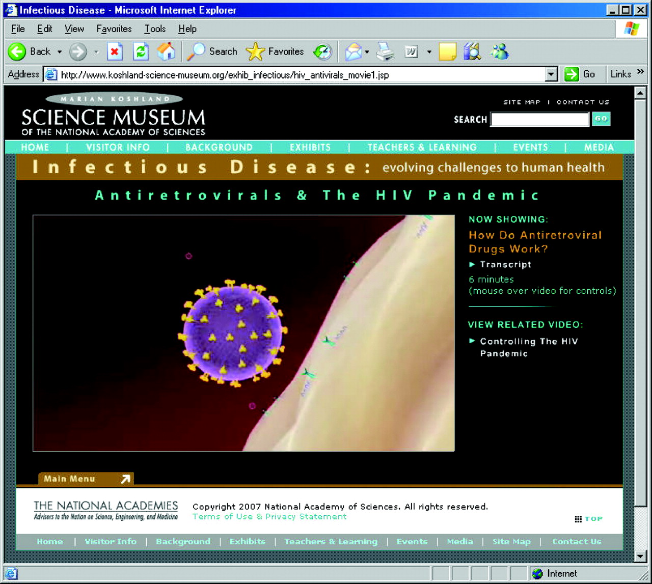
Figure 6. The Koshland Science Museum of the National Academy of Sciences has a good animation of the HIV life cycle that emphasizes drug targets.
Viral Informatics
The accumulation of data on viral genomes makes it increasingly possible to identify and classify viruses by the content of their genome, by simply reading sequence. When the AIDS epidemic surfaced in 1981, such information and technologies were not available, and understanding AIDS was delayed. Today, rapid sequencing technology and growing sequence databases have helped to identify a strong candidate virus responsible for the devastating Colony Collapse Disorder in honeybees. The Koshland Science Museum has an excellent Flash-based activity that demonstrates how DNA sequence was used to classify the previously unknown virus responsible for the emerging Severe Acute Respiratory Syndrome (SARS) epidemic in 2002 (http://www.koshland-science-museum.org/exhibitdna/inf01.jsp#). To begin, you are given a sequence from the virus isolated from a SARS patient. You have the option of scrolling through 11,000 viral sequences trolling for a match, or you can tell the computer to find a match for you, a good lesson on the role of computing in bioinformatics. The feature then introduces the virus DNA chip, containing the sequences of all known viral genomes. The user is led through standard DNA microarray technology and methods, but in the analysis section a new twist is introduced, one implemented by David Wang, Joe DeRisi, and Don Ganem. Their clever innovation of converting a 2D matrix into a 1D “barcode” streamlines the search for a viral match by condensing the most relevant genomic information, while also making it easier for human eyes to interpret and manipulate. The best sequence match in the case of the Virus 3 sample, from a SARS patient, is close but imperfect, consistent with SARS being caused by a previously unidentified virus in the family of coronaviruses.
Although viral genomes are small and simple compared with the genomes of cellular organisms, their diversity, sheer number, and overlapping reading frames present specific sequencing, alignment, and annotation challenges. The Microbiology Bytes website has a good overview of viral genomes (http://www.microbiologybytes.com/introduction/genomes.html), including a useful chart comparing the size ranges of various genomes. Viral genomes can be explored on the National Center for Biotechnology Information website (http://www.ncbi.nlm.nih.gov/genomes/VIRUSES/viruses.html) using the Basic Local Alignment Search Tool (BLAST) and related applications. For many reasons, a lot of energy has gone into the development of informatics software that extends or offers alternatives to BLAST. The Viral Bioinformatics Resource Center (http://athena.bioc.uvic.ca/) at the University of Victoria has developed what looks like a very useful suite of tools for analyzing and comparing diverse viral genomes. The website is well organized, with clear graphics and descriptions of their database structure and associated sorting and analysis tools. I was also impressed that the site featured a good tutorial and is supported by an online forum and a blog by Chris Upton.
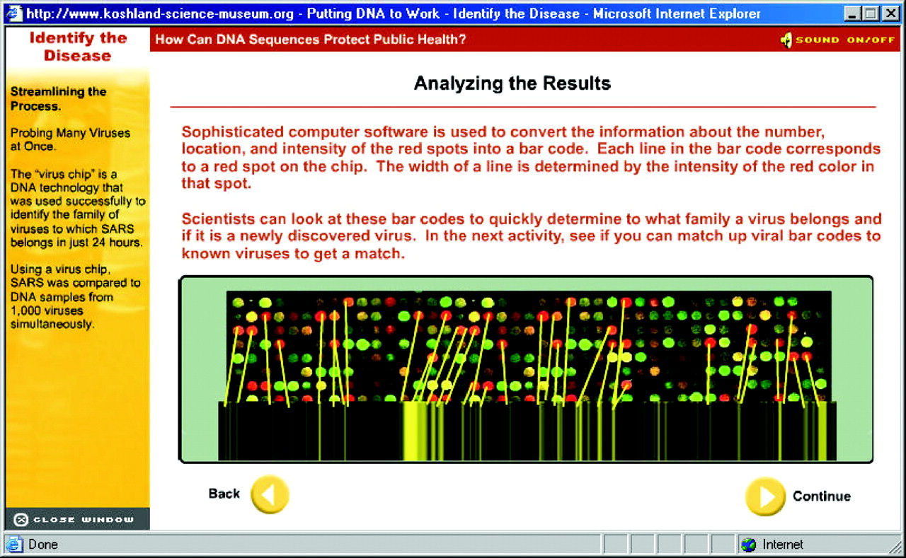
Figure 7. The Koshland Science Museum website has an excellent activity showing how genomic information can be used to identify new disease agents in emerging epidemics, in this case SARS.
We are virtually awash in a world of unidentified viruses, each with their own unique genome, which presents a terrific opportunity to combine authentic scientific discovery with education. The HHMI is launching a new Science Education Alliance (SEA) (http://www.hhmi.org/bulletin/aug2007/chronicle/sea.html and www.hhmi.org/grants/sea) aiming to foster innovation in science education. One of the first SEA activities will be to develop a national genomics experiment centered on isolating mycobacterial phage and sequencing their genomes. The project is inspired by the work of Graham Hatfill (http://www.pitt.edu/∼gfh/), Sally Elgin (http://www.nslc.wustl.edu/elgin/genomics/index.html), and others working with undergraduate students. The project promises to yield important information regarding viral ecology and evolution. A visit to the University of Leicester's Introduction to Molecular Virology (http://www.mcb.uct.ac.za/tutorial) should convince students that there is much to be learned in the area of viral origins (http://www.mcb.uct.ac.za/tutorial/virorig.html). Another website that presents a good outline of viral evolution can be found at EvoWiki.org (http://wiki.cotch.net/index.php/Main_Page), which focuses on the evolution and creationism controversy and has lots of good information on evolutionary biology, including a good section on viral origins and evolution (http://wiki.cotch.net/index.php/Virus).
Viral Bestiary
The life cycle of the double-stranded DNA herpes simplex virus, well-animated on the Sumanas website (http://www.sumanasinc.com/webcontent/anisamples/microbiology/herpessimplex.html), is fascinating. However, a student is unlikely to ever forget an actual picture of the herpes virus. You can see its unmistakable fried egg appearance on the webpages of Dr. Linda Stannard of the University of Capetown (http://web.uct.ac.za/depts/mmi/stannard/herpes.html). Her Virus Ultra Structure website (http://web.uct.ac.za/depts/mmi/stannard/virarch.html) has a very good description of viral architecture including some history of the field. The site has a particularly strong collection of electron microscope images, and Dr. Stannard's online lecture notes are a wealth of good information. The University of Wisconsin's Virus World (http://www.virology.wisc.edu/virusworld/) is another website rich with images and models of various viruses.
One of the more comprehensive websites on viruses is All the Virology on the WWW at www.virology.net, aiming to be the single most comprehensive site for virology on the Internet. Although the site contains general information on viruses, the images are its strong suit. One of the site's best features is The Big Picture Book of Viruses, where you can find images of viruses by family, individual names, structure, genome type, host, disease, etc. If you look up influenza alphabetically, you get a page on the family Orthomyxoviridae, with good photos of various flu viruses. There's also information on the genome and morphology and links to related websites. I've been told that the number of viral types is essentially infinite and the list of viruses in the Big Picture Book, while finite, is impressively long. The very first virus on the alphabetical list makes an important point about viral adaptability. Abadina virus, in the family Reoviridae, is able to infect invertebrate, plant, and vertebrate hosts. The site lists ∼70 families of viruses. The categories of hosts number 10: algae, archaea, bacteria, fungi, invertebrates, Mycoplasma, plants, protozoa, Sprioplasma, and vertebrates. Unfortunately, some links are in need of updating; for example, none of the links to various portions of the National Library of Medicine website are current.
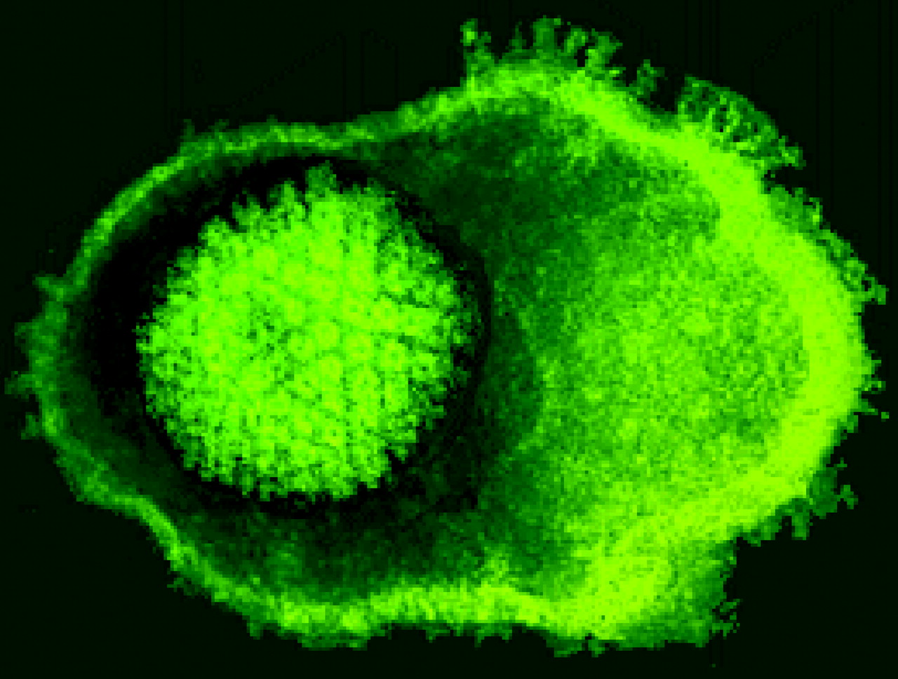
Figure 8. The unforgettable fried-egg appearance of herpes simplex virus, as seen on the Virus Ultra Structure website, where images of many different viruses can be found.
The elucidation of viral structures has progressed beyond flat transmission electron micrographs (TEMs). Crystallographic and other techniques for elucidating molecular structures have made it possible to build accurate and beautiful 3D models of viruses. Because most viral capsids contain millions of atoms, molecular graphics packages that work with Protein Data Bank files don't perform well for many viruses. Tom Goddard, Conrad Huang, and Thomas Ferrin of the University of California, San Francisco (UCSF) have published a beautiful online poster that discusses the issues involving good 3D modeling of large molecular structures like viruses. Although the poster (http://www.cgl.ucsf.edu/Research/virus/poster/poster.html) dates from 2004 and is not a full-fledged website, it is worth visiting for the stunning graphics. The gallery of 100 viral capsid images is an invaluable glimpse of viral diversity. These sort of high-end online “posters” are an interesting trend that greatly enhances the reach of traditional poster sessions. To learn more about the current state of the authors' software, you can visit the UCSF Chimera website (http://www.rbvi.ucsf.edu/chimera/) or view a recent online posting from Tom Goddard at http://www.cgl.ucsf.edu/chimera/assemblies06/assemblies.html. An alternative set of tools (http://viperdb.scripps.edu/first_time.php), developed by The Scripps Research Institute, is focused on imaging icosahedral (20-sided polyhedron) viral structures entered in the Protein Data Bank.
Thwarting Viruses
Although one of the best-known means for fighting viruses is an effective vaccine, I've found a paucity of websites aimed at education on the science of vaccine development. The Microbiology Bytes website has a lot of good information in the virus section, including a description of how vaccines work (http://www.microbiologybytes.com/virology/Vaccines.html). A South African–based animator has made a very engaging animation about an HIV vaccine (http://blog.iaminawe.com/2007/07/03/hiv-vaccine-animation/). The audience for the animation is the general public, but the scientific basics are present, and the colloquial narration is a refreshing antidote to the typical corporate-produced animations. In addition to the immune system, cells have another defense against viral invaders—the mechanism of RNA interference (RNAi). The use of RNAi as a research tool has somewhat overshadowed its hypothesized evolution as a means to thwart viruses and other disruptive mobile genetic elements. The NOVA ScienceNow website has an excellent interactive feature on RNAi. Go to http://www.pbs.org/wgbh/nova/sciencenow/3210/02.html and click on “RNAi Explained.” The feature makes use of engaging character animation and a clever metaphor. In many ways the animation is more effective in the original context of the 15-min television segment, which can be viewed by clicking on “Watch the Segment.”
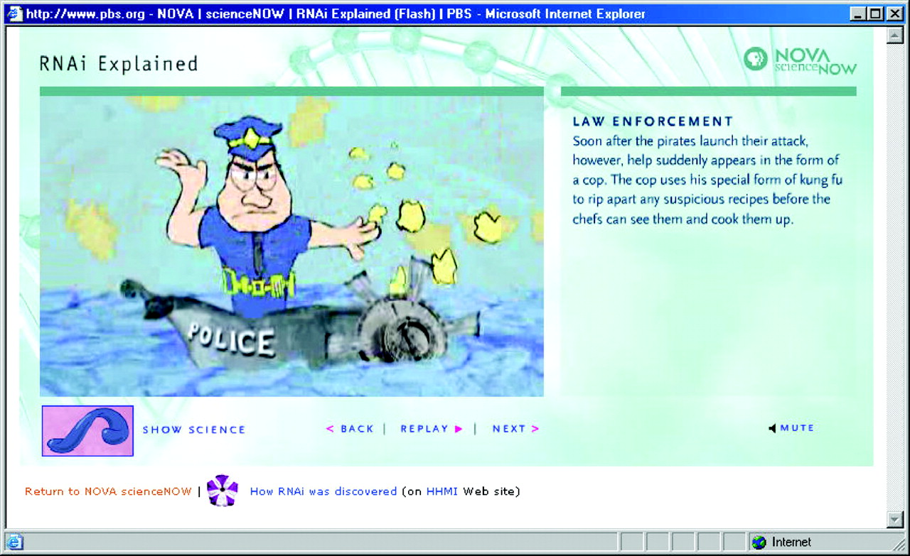
Figure 9. The NOVA ScienceNow webpages have a very engaging animated metaphor for explaining RNA interference.
As always, your comments are appreciated and can be directed to the author at [email protected].


