Using a Replica of Leeuwenhoek's Microscope to Teach the History of Science and to Motivate Students to Discover the Vision and the Contributions of the First Microscopists
Abstract
The history of science should be incorporated into science teaching as a means of improving learning and also to increase the students' understanding about the nature of science. In biology education, the history of microscopy deserves a special place. The discovery of this instrument not only opened a new and fantastic microworld but also led to the development of one unifying principle of biological sciences (i.e., cell theory). The microscopes of Leeuwenhoek and Hooke opened windows into the microworld of living organisms. In the present work, the knowledge of these themes was analyzed in a group of students beginning an undergraduate biology course. Our data suggest that the history of microscopy is poorly treated at the secondary school level. We propose a didactic activity using a replica of Leeuwenhoek's microscope made with Plexiglas and a lens obtained from a key chain laser pointer or from a broken CD drive. The proposed activity motivated students to learn about microscopy and helped them to appreciate scientific knowledge from a historical perspective.
INTRODUCTION
“… were incredibly small, nay so small, in my sight, that I judged that even if 100 of these very wee animals lay stretched out one against another, they could not reach to the length of a grain of coarse sand. ” (Leeuwenhoek, 1666)
Scholars have recognized that the history of science should be included in the science curriculum, both at the secondary and university level (Matthews, 2004) to improve the students' conceptions about the nature of science (NOS). In fact, over the past decade, the NOS has enjoyed renewed attention among science educators as a principal component of scientific literacy (National Research Council, 1996). Recent studies specify that science teachers should not only teach in a manner consistent with current views of the scientific enterprise but also purposefully instruct students in specific aspects of the NOS. To improve this situation, many researchers have recommended initiatives such as teaching about the history of science to help students develop more accurate views about the NOS (Duschl, 1990; Matthews, 1994; Hsu and Lee, 1995; Monk and Osborne, 1997). Studying the history and the philosophy of science develops a better understanding of the nature of the scientific enterprise and an appreciation for how science concepts change with the time. It also leads to a better understanding of the concepts themselves (i.e., it improves science learning). The history of science provides contextual information of what definitions, thoughts, concepts, and theories of science have prevailed during different moments in history. Also, it shows that science is a human endeavor, and it reveals the deficiencies of such a human effort (Dass, 2005).
In biology teaching, it is important to call attention to the contributions made by the first microscopists during the seventeenth century, who described a completely new world. In fact, these investigators first demonstrated the existence of microorganisms present in a drop of water or vinegar, and they also gave detailed descriptions of many tiny structures such as insect and plant parts.
One pioneer in the use of the microscope was Robert Hooke (1635–1702), an English physicist. In 1665, Hooke published Micrographia that described not only minute structures but also distant planetary bodies, the Wave theory of light, the organic origin of fossils, and various other philosophical and scientific subjects. Hooke coined the term “cells” to refer to the units that he saw in cork slices. Cella is a Latin word meaning “a small room” that monks inhabited. Latin-speaking people applied the word cellulae to the six-sided cells of the honeycomb. Part of this book can be accessed online at http://archive.nlm.nih.gov/proj/ttp/flash/hooke/hooke.html.
Another prominent microscopist of the seventeenth century was Antony van Leeuwenhoek (1632–1723), a tradesman from Delft, Holland. The reading of Hooke's book is believed to have roused an interest in van Leeuwenhoek to use the microscope to investigate the natural world. He became an expert in constructing extremely simple microscopes using only one lens, mounted in a tiny hole in the brass plate that made up the body of the instrument. Using these simple devices, he was the first to observe and describe single-celled microorganisms, which he originally referred to as animalcules. He was also the first to record microscopic observations of muscle fibers, bacteria, and spermatozoa. In a long series of papers presented to the Royal Society of London, he described many specific forms of these microorganisms and structures. Throughout his lifetime, van Leeuwenhoek made >400 different microscopes, but only a dozen of these still exist today. Those that have survived are able to magnify up to 275 times. However, it is suspected that van Leeuwenhoek possessed some microscopes that could magnify up to 500 times.
The descriptions of these first microscopists were crucial to the subsequent development of biological theories in the succeeding centuries. For example, it permitted the refutation of the theory of spontaneous generation by Louis Pasteur (1822–1895) two centuries later, as well as the refutation of the miasma disease theories (Kahtan and Greenberg, 1992). The development of cell theory (Schleiden, 1804–1881; Schwann, 1810–1882), which is one of the most important theories in biology, was a consequence of these pioneering works ( Mazzarello, 1999).
In Brazil, the textbooks for the first year of secondary level biology always show figures of the Leeuwenhoek and Hooke microscopes and describe briefly the history of first microscopic observations and the genesis of the cell theory. Despite this exposure, our experience has shown that students arrive at university without basic knowledge of these historical facts. Many students are unable to recognize the Leeuwenhoek device as a microscope, even though this figure was presented in their secondary school textbooks. Our experience also has shown that traditional ways of teaching these historical facts, such as a text reading, do not normally promote enthusiasm in the students.
We are living in the information era, and the Internet has revolutionized the means and the rate at which we can obtain information. More than just transmitting information, our didactic activities aim to promote curiosity and to motivate students to independently acquire knowledge.
The objectives of this work were to 1) analyze what undergraduate biology students remembered about the history of the first microscopists from their regular biology courses in high school; 2) quantify the fraction of students who are able to recognize the Leeuwenhoek microscope; and 3) propose and test a didactic activity involving the history of biology that is capable of motivating “self-learning.”
PROPOSITION OF A DIDACTIC ACTIVITY TO DEVELOP INTEREST IN THE HISTORY OF BIOLOGY
Study Participants
The participants of this study were undergraduate students at the start of the bachelor-level course in biological sciences at Santa Maria University in Brazil. Our sample consisted of 132 students who were enrolled in the first-semester cell biology course. Two classes were included in this study, and they were named the 2008 class and the 2009 class.
Class Activities
The class activities were very simple and quick, consisting of four parts or steps. First was the presentation of some questions to verify the level of information about history of science. This was followed by practical activities to create a sensible view of the question that was then followed by a challenge task. Finally, an evaluation was used to access the level of motivation created and determine whether the students could remember these activities and related information for a long time.
In the first step, the pretest was applied. The pretest was composed of multiple-choice questions that were designed to verify whether the students correctly associated pictures, names, and dates about the history of the microscope. At least three questions regarding the same subject were presented, and a student was only classified as “knowing the information” if he or she answered correctly the whole set of questions.
Afterward, during the sensibilization step, the students were invited to use a microscope similar to Leeuwenhoek's. The rationale for this moment was to promote curiosity about the early microscopes and contributions of the first microscopists. Later, the students were encouraged to search for information about early microscopes and microscopists themselves. These class activities are detailed below.
First Step: Destabilization. Promoting situations in which our knowledge should be put in check is sometimes a good way to become disposed to learn more about a subject. Using this principle, we prepared a set of questions in a PowerPoint presentation for the students, and they received a grade to record their choices. Two groups of questions were presented. In the first, the students were asked whether the microscope had already been invented in relation to other historical facts, such as the invention of the electric lamp, the first pox vaccine application, Gutenberg's first book printing, and the arrival of Columbus in the Americas. The goal of these inquires was to see whether the students were able to roughly identify the historical period of microscope invention.
The purpose of the second query set was to see whether the students were able to identify the first microscopes, and the set consisted of figures of devices that were to be identified as microscopes or not microscopes. Eighteen figures were used. Some pictures corresponded to the first microscopes, made by Leeuwenhoek and Hooke, and others were pictures of modern optical and electronic microscopes. Other equipment, such as telescopes, polarimeters, and spectrophotometers, also was shown.
In the other part of the test, the students responded freely to the following three questions: 1) When were microorganisms seen for first time, and who described them? 2) What do they imagine the first microscopes were like? 3) What is the cell theory and who formulated it?
Second Step: Sensibilization. In this step, the students had the chance to use a “replica” of Leeuwenhoek's microscope and to see different microscopic structures such as onion cells, Paramecium and other microorganisms, insects, and parts of plants. The goal of these activities was to allow the students to observe that a very simple device can permit a meaningful observation of the “microworld.” Also, this activity served to highlight how marvelous the discovery of this microworld would have been centuries ago.
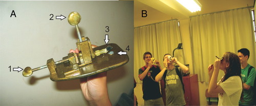
In the literature, there are some wonderful descriptions on how to construct a replica of Leeuwenhoek's microscope, including the website at www.mindspring.com/∼alshinn/Leeuwenhoekplans.html. However, these replicas are not simply made. We have developed a simpler device to be used in our classes. The lens is obtained from a key chain laser pointer or from a broken CD drive. The microscope body is made of acrylic glass (Plexiglas), a screw, and epoxy putty that have been painted with bronze paint (Figure 1A). Another even simpler device can be made using recycled material such as a polyethylene terephthalate bottle and a plastic box. The description of how both apparatuses can be constructed can be found in the Supplemental Material.

Figure 1. (A) A view of Leeuwenhoek's replica microscope made with Plexiglas and a lens obtained from a key chain laser pointer. 1, “control stage” screw; 2, focus screw; 3, “slide” with preparation; and 4, lens. (B) Students in a classroom making observations with the replica of Leeuwenhoek's microscope.
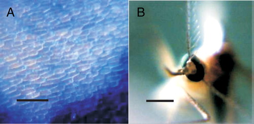
Figure 2depicts some materials, such as onion cells and mosquitoes, that were observed by the students using the replica of Leeuwenhoek's microscope. These photos were obtained directly by the homemade microscope by using a webcam. The microscopes made using a chain laser pointer lens are able to magnify approximately 80–100 times, whereas using a CD lens magnifies approximately 200 times. This last magnification is approximately the same as that of the Leeuwenhoek microscope. These activities do not need to be performed in the laboratory; we conducted them in the classroom. Different materials such as microorganisms, plant tissues, and insects were put in various microscopes, and the students shared their observations and impressions (Figure 1B).

Figure 2. Materials observed in the Leeuwenhoek replica microscope. (A) Onion cells. (B) Mosquito. Bars, approximately 200 μm.
Third Step: The Challenge. During the sensibilization moment, the students were informed that the simple microscopes they were using corresponded to replicas of Leeuwenhoek's microscope. They were also told that in the same historical period, another scientist named Robert Hooke constructed a different microscope and made other important contributions. In addition, it was emphasized that the works of these first microscopists were essential to the development of biology.
We proposed to the students that they search for more information about the history of microscopy and early microscopists by using the Internet. We also suggested that they describe their personal experience with these class activities. One group was informed that they could send the results of their Internet searches to the teachers via email (2008 class), and the other group did not receive this suggestion (2009 class). It was clear to both groups that this Internet search was a voluntary activity that would have no bearing on the students' grades.
Fourth Step: Evaluation. The effectiveness of these class activities in promoting learning, as well as the students' motivation to perform an Internet search about the history of the microscope, were evaluated by comparing pretests and posttests (Sundberg, 2002). These two categories were assessed by using five groups of questions presented to the students at two different time points: 3 wk after the practical activities (2009 class) and 16 mo later (2008 class). This temporal and spatial separation between the evaluations given to the two student groups allowed for identification of not only short-term learning but also long-term retention of knowledge. The average scores of both groups in pre- and posttest questions were compared using t test.
Only one posttest question was “open-ended”; it asked whether the voluntary search had been conducted and why. The other posttest questions asked for information that should have been obtained during the Internet search (not presented during class). The posttest also permitted an evaluation of what conditions were more effective in motivating the students and promoting learning: asking for written reports (2008 class) or simply suggesting that the students seek additional information by using the Internet (2009 class).
RESULTS AND DISCUSSION
What Did the Students Know about the History of the First Microscopists? The Pretest Results
The studied sample was composed of two groups of students (2008 class and 2009 class). The same pretest was administered to both groups at different times: for the 2008 class, the pretest was administered in October 2007; for the 2009 class, it was administered in April 2009. The scores for both groups were compared, and they were very similar. No statistically significant difference in pretest answers was found between the 2008 class and 2009 class.
Only 19.3% of our students were able to recognize the Leeuwenhoek microscope, even though modern optical microscopes were recognized (Table 1). Other early equipment, such as that made by the Jansens, was only recognized by 25.4% of students. However, Hooke's device was identified as a microscope by 77.3% of the students, probably because it is a compound microscope that more closely resembles a modern microscope. The modern optical microscope, with an ocular, nose piece, and stage, is a symbolic scientific instrument and was recognized well by the students. However, the history of this apparatus and its role in the development of biological theories are not recognized extensively.
| Pretestresultsa | Posttest resultsa | |
|---|---|---|
| Historical period of firstmicroscopists | 10.2 | 58.33 |
| Leeuwenhoek's microscope | 19.3 | 90.91 |
| Hooke's microscope | 77.3 | 90.91 |
| Jansen's microscope | 25.4 | 90.91 |
| Recognize the firstmicroscopists | —b | 87.88 |
| Biological material analyzedby the first microscopists(Micrographia pictures) | 21.5% | 54.54 |
| Modern optical microscope | 100 | No question included |
| Electron microscope | 97.7 | No question included |
| Other equipment (e.g.,spectrophotometer,polarimeter) | 85 | No question included |
The historical period in which Leeuwenhoek and Hooke lived and made their contributions is also not known by the majority of students (89.8%). Even Robert Hooke's name was only remembered by three students.
Our data show that students entering university in Brazil have not learned the history of the discovery of the microworld or the genesis of cell theory, despite the fact that secondary school textbooks contain these topics.
Developing Interest in the History of Science
Mentioning to the students that their teachers would like to receive the results of their Internet searches about the history of microscopy was very effective in mobilizing the students to perform the search (2008 class). The majority of students (96.5%) spontaneously performed a search and sent their report to the cell biology course website to be shared with classmates. Analysis of the posttest revealed that the students' answers could be separated into two subgroups. One subgroup (52.3%) said that the reason for performing the suggested search was curiosity about the topic. The other subgroup (47.7%) responded that the reason was the suggestion made by the teachers.
The data gathered from students in the 2009 class, who did not receive a suggestion to send search reports to their teachers, were satisfactory, although a low percentage (65.8%) of students responded that the search had been performed. The major reasons given by the students for not completing the suggested activity were as follows: “I had problems with Internet access at home” and “I forgot about it.” It is remarkable that a majority of the students (52.3% in the 2008 class and 65.8% in the 2009 class) answered in the posttest that they performed the voluntary search and cited curiosity as the reason.
Also, the personal experience descriptions about the class activities were heartening; the majority of the students evaluated the activities very positively. As an illustration, we describe some reports below.
Student 1. I am sure it was a unique experience, never have I imagined that so small and simple a microscope could be so interesting… While today is well known that many “animals” exist that cannot be seen without the help of a lens, even now it is curious to see these creatures moving. I loved to see the “microbes” moving in the water, the onion cells, as well as some details in the fruit fly. It is marvelous to know that with a so simple microscope, it is possible to see a lot of things ….
The opportunity to see a “replica” of the Leeuwenhoek microscope was marvelous, and I can say that I loved this class. I did not have an idea that we could make such a simple microscope and that it would work so well.
Student 2. I didn't know the microscope history. I was enchanted when I could use the replica made by the teacher. Also, I could see after in the Web search that the replica is much like the original.
Student 3. I never imagined that in my first cell biology class I would be using a replica of the first microscope that is so different from those we normally see. It was a surprise to me to see the simplicity of this device and also Leeuwenhoek's great creativity in making it. Also, it is interesting to observe the same difficulties that the first microscopists had, such as the problems with the luminosity and obtaining the focus.
Effectively Learning the History of Microscopy
The same posttest was applied to both groups in April 2009 (more than a year after the pretest for the 2008 class; 3 wk after the pretest for the 2009 class). We did not find differences in the provided results between students from the 2008 and 2009 classes.
The activities conducted resulted in effective learning. The students' capacities to recognize the Leeuwenhoek and Hooke microscopes increased significantly (Table 1). In the pretest, only 19.3% of the students correctly identified the picture of Leeuwenhoek's microscope; after the educational activities, 90.91% of them were able to do so. These results indicate that the activities did not simply promote short-term learning but in addition were very effective in promoting long-term retention.
Similarly, correct identification of the historical period in which the first microscopists made their contributions had a significant improvement. In the pretest, only 10.2% of the students were able to identify the correct time period, whereas in the posttest 58.33% of students correctly identified this historical period.
The posttest results also indicated that the search for more information about the history of microscopy and early microscopists by using the Internet was effective. Approximately 54% of the students were able to recognize pictures presented in Micrographia and associated them with Hook's name in multiple-choice questions. This information was not included in the activities; and, in the pretest, the students had a very low percentage of correct answers to the questions about it (Table 1).
The consistency of the posttest answers was confirmed by “triangulation.” The students who correctly answered the questions regarding the historical period during which the microscope was invented and the names of the first microscopists were the same students who stated that they conducted the voluntary search.
CONCLUSIONS
The main conclusions that can be obtained from our work are as follows: 1) the invention of the microscope and the discovery of the microworld by Hooke and Leeuwenhoek is greatly ignored by secondary students; 2) the role of the invention of the microscope in the history of biology is also disregarded; 3) making observations with a replica of the Leeuwenhoek microscope is a highly motivating didactic activity; 4) the simple activities described here can promote students to learn more about the history of science and may enable students to look at the knowledge with a historical perspective; and 5) these activities are effective in the learning of microscopy history.
Wang and Marsh (2002) pointed out that the history of science is poorly addressed in high school and even at the undergraduate level, and our results agree with this observation. One disturbing aspect of our study is that even students who have selected to study biology had a limited knowledge about one of the unifying principles of biological sciences (cell theory), and they also had only vague ideas about the discoveries that were important for the development of this principle. Considering that in Brazil only ∼5% of the students enter university, we can suppose that the familiarity with this fundamental aspect of biological science is almost absent in the entire population.
According to Gooday et al. (2008), “there are at least two ways in which science education needs the history of science”: 1) including history of science as part of the science curriculum; and (2) using it strategically to defend the autonomy of the science curriculum from inappropriate extrinsic forces, helping to produce a stronger training for those whose scientific careers will be forged in our schools, colleges, and universities in decades to come. We propose that simple activities, such as those described here, should be incorporated in the high school curriculum as an alternative strategy to increase students' motivation for learning biology on more solid ground. Furthermore, in addition to being a fun and highly motivating experience, including the history of the early microscopists and details of how their discoveries contributed to the development of cell theory in the regular high school curriculum will probably contribute to increasing the students' perceptions about the real nature of science.


