Explain the Brain: Websites to Help Scientists Teach Neuroscience to the General Public
INTRODUCTION
The field of neuroscience has experienced enormous growth over the past few decades. The Society for Neuroscience, founded in 1969 with only 500 members, now has ∼42,000 members. Not only are research findings from neuroscience laboratories around the world continuing to influence biology and medicine, the public is becoming increasingly aware of the importance of brain research in their lives. Educators look to neuroscience to become better teachers, lawyers and judges explore the literature to gain insight into court cases, and marketers consider the use of brain scans to glean information about consumer preferences. The importance of neuroscience is further reinforced by the recent funding of large-scale programs to support brain research, such as the BRAIN Initiative (www.nih.gov/science/brain/index.htm) and the Human Brain Project (www.humanbrainproject.eu). In addition to supporting research, these initiatives encourage scientists to engage with the public with the goal of increasing people's understanding of neuroscience.
With this increased interest in brain research, neuroscientists are often invited to share their work with lay audiences. For example, Brain Awareness Week is an international event occurring each March that is dedicated to promoting the public and personal benefits of brain research (Burton, 2007; McNerney et al., 2009). Other medical, health, and science observations related to neural function are recognized at other times of the year, including World Meningitis Day, National Sleep Awareness Week, National Autism Awareness Month, Healthy Vision Month, Mental Health Month, National Aphasia Awareness Month, National Traumatic Brain Injury Awareness Month, and World Alzheimer's Month.
This Feature showcases Internet-based resources to help neuroscientists explain their work to the general public and K–12 audiences. These Internet resources feature videos, images, animations, and other outreach tools that are accessible to lay audiences and that speakers can adapt for their own presentations.
HISTORY OF NEUROSCIENCE
Tracing the historical foundations of neuroscience is one way to spark an audience's interest in this topic. The stories behind the significant events, discoveries, and important people shaping the field of neuroscience help to make this scientific topic more accessible and personal. For a timeline with links to other detailed resources, turn to the Neuroscience for Kids Milestones in Neuroscience Research site (http://faculty.washington.edu/chudler/hist.html). The Neuroscience for Kids timeline is illustrated with images from the Moody Medical Library (http://arweb5.utmb.edu/ar/Library/BlockerCollections/tabid/183/Default.aspx) and the U.S. National Library of Medicine (www.nlm.nih.gov/hmd/index.html) that can also be used when creating a historical presentation (see Figure 1).
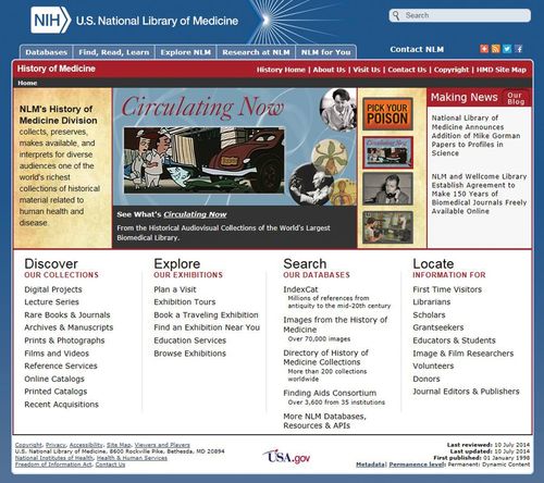
Figure 1. History of Medicine page within the U.S. National Library of Medicine website (www.nlm.nih.gov/hmd/index.html). Navigate to “Images from the History of Medicine” to locate photographs, portraits, and drawings about the history of brain research. The screenshot image is courtesy of the National Library of Medicine.
Researchers who study specific neurological diseases and disorders may want to visit Founders of Neurology (www.uic.edu/depts/mcne/homepage/neurofounders.html) for pictures of the early contributors to clinical neuroscience and short summaries of their work. Who Named It? (www.whonamedit.com) provides a list of medical eponyms, searchable alphabetically or by disease name, with information about the person for whom each disease is named.
Significant discoveries in neuroscience can be highlighted by sharing the biographies of the researchers who have received a Nobel Prize in Physiology or Medicine. The official Nobel Prize site (www.nobelprize.org) features biographies of these prize-winning researchers and descriptions of their groundbreaking research. By navigating to the page announcing Nobel Prize laureates in physiology or medicine (www.nobelprize.org/nobel_prizes/medicine/laureates), users can learn about Camillo Golgi and Santiago Ramón y Cajal (1906 laureates); Sir Charles Scott Sherrington and Edgar Douglas Adrian (1932 laureates); Sir John Carew Eccles, Alan Lloyd Hodgkin, and Andrew Fielding Huxley (1963 laureates); Arvid Carlsson, Paul Greengard, and Eric R. Kandel (2000 laureates); and many other pioneers in the field of neuroscience.
Presenters with an interest in neurology, psychiatry, and neurosurgery can find primary source documents and images from the U.S. National Library of Medicine's Diseases of the Mind: Highlights of American Psychiatry through 1900 (www.nlm.nih.gov/hmd/diseases/index.html) and the Cyber Museum of Neurosurgery (www.neurosurgery.org/cybermuseum/summary.html).
When scientists give public lectures, they will likely encounter people who are familiar with the clinical cases of Phineas Gage and Henry Molaison. Gage suffered a traumatic brain injury when an iron tamping rod went through his head in 1848, and Molaison suffered from amnesia after he had brain surgery to control seizures in 1953. The life of Phineas Gage and subsequent research to understand his symptoms are documented by Dr. Malcolm Macmillan on his Phineas Gage Information page (www.uakron.edu/gage). The Brain Observatory (http://thebrainobservatory.ucsd.edu/hm) at the University of California, San Diego, details Molaison's life and contains an atlas of his brain reconstructed from histological examination of 2401 thin-tissue slices.
NEUROANATOMY
The complex structure of the brain, while fascinating, is often difficult for young students to grasp. Although many students come to understand the three-dimensional organization of the brain through hands-on dissection and examination of the brain, not all presenters have access to brain specimens. The Internet brings neuroanatomical specimens out of the lab and onto the computer, where presenters can move through images of the brain at their own pace as they describe brain structure. Such displays reduce safety concerns associated with handling brain specimens (Budka et al., 1995) and allow presenters to share their knowledge of neuroanatomy with larger audiences.
Comparative neuroanatomy of animal species is one way to engage students in the study of brain structures. The Comparative Mammalian Brain Collections (http://brainmuseum.org;Figure 2) contains a collection of photographs from nearly 200 different species. Many of the specimens displayed on the website have been stained (cells and/or fibers) and photographed to provide an excellent resource for comparative neuroanatomy. High-resolution images of human, monkey, wolf, cat, rat, mouse, opossum, goldfish, and chicken brains can be manipulated using BrainMaps.org (http://brainmaps.org). As an adjunct to a hands-on sheep brain dissection, Dr. Timothy Cannon and his colleagues have created the Sheep Brain Dissection Guide (www.scranton.edu/faculty/cannon/sheep). The newest version of this guide will assist presenters working with students at the elementary, high school, and college levels.
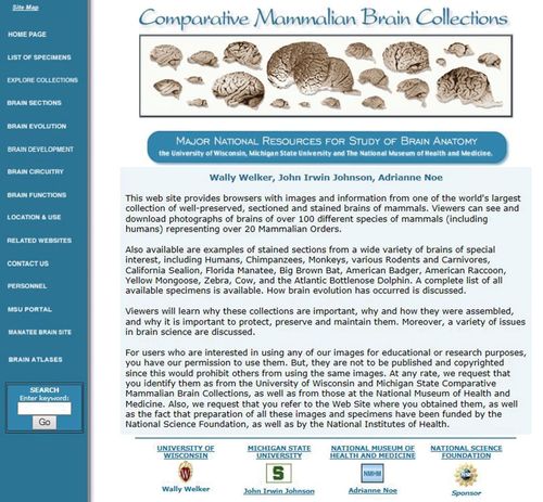
Figure 2. The Comparative Mammalian Brain Collections (http://brainmuseum.org) contains photographs of more than 100 different animal brains. This screenshot image is reproduced with permission from www.brains.rad.msu.edu and http://brainmuseum.org, supported by the U.S. National Science Foundation.
Many online resources are available to assist presenters in explaining human brain anatomy. One example is the interactive atlas of the human spinal cord and brain stem neuroanatomy developed by University of Louisville Physical Therapy (www.bellarmine.edu/faculty/mwiegand/atlas/cover.html). This site allows users to move through 27 sections from the base of the spinal cord to the thalamus. Each plate illustrates the level of the section and labels major structures. The U.S. National Library of Medicine's Visible Human Project (www.nlm.nih.gov/research/visible/visible_human.html) was used to create another neuroanatomical “fly-through” of the human brain called LUMEN Cross-Section Tutorial (www.meddean.luc.edu/lumen/meded/grossanatomy/x_sec/mainx_sec.htm). LUMEN illustrates the brain and spinal cord through 12 horizontal sections, using photographs, magnetic resonance images (MRI), and computer tomography (CT). HeadNeckBrainSpine (http://headneckbrainspine.com) also uses MRI and CT. The Whole Brain Atlas (www.med.harvard.edu/AANLIB/home.html) uses positron emission tomography (PET) and single-photon emission computed tomography, in addition to MRI and CT, to illustrate normal and abnormal brain anatomy in the horizontal, sagittal, and coronal planes. Nissl- and fiber-stained human brain sections in these three planes are also featured in the aptly named Zoomable Human Brain Atlas (http://zoomablebrain.bio.uci.edu;Figure 3).
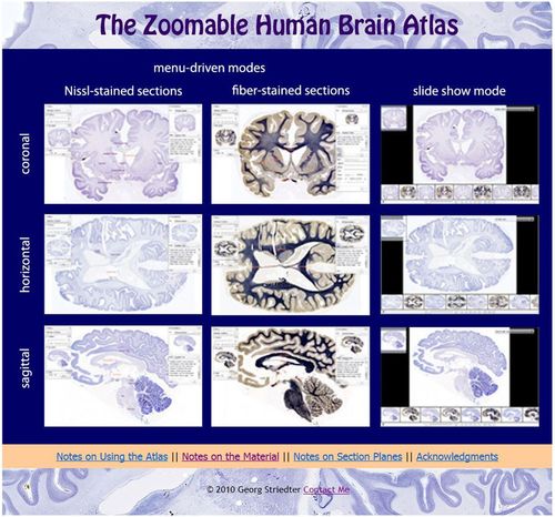
Figure 3. The Zoomable Human Brain Atlas website (http://zoomablebrain.bio.uci.edu) contains Nissl- and fiber-stained sections of the human brain in sagittal, horizontal, and coronal sections. This screenshot image is used with the permission of Georg Striedter, PhD, Department of Neurobiology and Behavior, University of California, Irvine.
Presenters, especially those who will be speaking in classrooms, may want to challenge students to create models of the brain and neurons. BrainU (http://brainu.org; see the Lessons>Model Building link) and Project NEURON (http://neuron.illinois.edu) provide examples of physical models, such as bead neurons and neuronal communication gloves, to teach concepts about brain anatomy, neuronal structure, and neurotransmission. Animations and videos illustrating these concepts can be located at Howard Hughes Medical Institute's BioInteractive site (www.hhmi.org/biointeractive/explore-neuroscience).
NEUROPHYSIOLOGY
The functioning of the nervous system can be explained to large and small audiences using animations, demonstrations, and games. Several textbooks, for example, Biological Psychology: An Introduction to Behavioral, Cognitive, and Clinical Neuroscience, 7th edition (Breedlove and Watson, 2013; http://7e.biopsychology.com/outline03.html) and Neuroscience (Purves et al., 2012; http://sites.sinauer.com/neuroscience5e) have companion websites with excellent animations that illustrate resting potential, action potential, and synaptic transmission. For young students, BrainU (http://brainu.org/movies) has created several cartoon-like animations about neurotransmission (Figure 4). To use animations, presenters will need to use newer Web browsers and to download multimedia applications such as Apple QuickTime, Adobe Shockwave, or Adobe Flash Player.
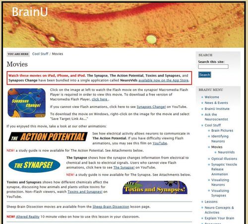
Figure 4. The BrainU website (http://brainu.org/movies) features several animations illustrating neurotransmission. This screenshot image is used with the permission of Dr. Janet Dubinsky, University of Minnesota.
THE SENSES
How much? Where? When? What? These are questions about the constant barrage of sensory information flooding into the nervous system that presenters can use to interact with audiences. Visual illusions printed on cards or projected onto a screen are easy to make and can effectively illustrate how the brain processes information from the outside world. Examples of visual illusions can be found at:
Optical Illusions & Visual Phenomena (www.michaelbach.de/ot): more than 100 different demonstrations, many interactive
Exploratorium (www.exploratorium.edu/explore/staff_picks/optical_illusions): interactive demonstrations with easy-to-understand explanations
Illusions Gallery (http://dragon.uml.edu/psych/illusion.html): simple visual illusions with explanations
Brusspup: Illusions and Science (www.youtube.com/brusspup): video channel featuring unique illusions
The Best Visual Illusion Contest (http://illusionoftheyear.com): award-winning illusions from an annual competition
Your Amazing Brain: Super Senses (www.youramazingbrain.org.uk): a collection of common visual illusions with explanations
Auditory illusions are more difficult to find online. The McGurk effect (http://auditoryneuroscience.com/McGurkEffect;http://epsych.msstate.edu/descriptive/crossModal/mcgurk/index.html), which shows how vision can influence what we hear, is an outstanding demonstration of an auditory illusion.
NEUROPHARMACOLOGY
The topic of neuropharmacology as it relates to drug abuse and addiction may be of great interest to students, especially because many school districts have classes in which these topics are taught at specific grade levels. Speakers in need of slides, posters, images, and basic information about the health effects and consequences of drug abuse and addiction should turn to the National Institute on Drug Abuse website (NIDA; www.nida.nih.gov). Of particular note on the NIDA website are “teaching packets” that contain slide decks in PowerPoint format. “The Brain & the Actions of Cocaine, Opiates, and Marijuana” packet is a set of five presentations that discuss basic neuroanatomy of the brain and spinal cord, pain, neurotransmission, and reward pathways (www.drugabuse.gov/publications/term/41/Presentations). Posters, cards, pamphlets, and curriculum supplements can be ordered for free through the website and distributed in classrooms or at outreach events.
Classroom activities to educate students about the effects of drugs can be accessed through the Web Adventures site created at Rice University (http://webadventures.rice.edu/index.html;Figure 5). Web Adventures features two online, interactive games (N-Squad and Reconstructors) that focus on substance abuse. In addition to these online games, the website has classroom activities that can be used during presentations. Similarly, Learn.Genetics from the University of Utah (http://learn.genetics.utah.edu/content/addiction) and the Pharmacology Education Partnership from Duke University Medical School (www.thepepproject.net) have online modules about substance abuse accompanied by classroom lessons that are appropriate for intermediate or secondary students. Designed especially for middle school classrooms, Sowing the Seeds of Neuroscience (www.neuroseeds.org) from the University of Washington was developed to motivate students to learn about neuroscience by investigating the neuroactive properties of medicinal plants and herbs. A series of classroom lesson plans, including wet labs and demonstrations, can be downloaded from the Sowing the Seeds of Neuroscience website.
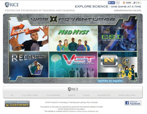
Figure 5. Web Adventures website (http://webadventures.rice.edu/index.html); the Reconstructors and N-Squad resources focus on neuroscience and neuropharmacology. This screenshot image is used with the permission of Dr. Leslie Miller, Center for Technology in Teaching and Learning, Rice University.
BEST PRACTICES
For scientists who usually teach students in graduate courses or who present their experimental results to their peers at a conference, speaking to young students and lay audiences may pose some unique challenges. Young students may have limited knowledge of the vocabulary used by speakers and no prior experience in neuroscience. By avoiding jargon and complex explanations, presenters who participate in outreach events have the opportunity to clear up common misconceptions about the brain (e.g., do we use only 10% of the brain; are people either right-brained or left-brained?). Professor Bruce Hood at the University of Bristol models excellent presentation skills in a video for the Royal Institution's 2011 Christmas Lecture (http://richannel.org/christmas-lectures/2011/meet-your-brain). Dr. Greg Gage also models effective speaking skills in a fun and engaging demonstration of the SpikerBox (Marzullo and Gage, 2012) during a TEDeD presentation (www.ted.com/talks/the_cockroach_beatbox). Other presentation tips can be found within the websites of the Society for Neuroscience (www.sfn.org/public-outreach/brain-awareness-week) and the Dana Foundation (www.dana.org/baw).
Looking for assistance with planning and executing an outreach event such as a school visit or a Brain Awareness Week activity? Graduate students who form the Brain Awareness Council at Wake Forest University (http://bac.graduate.wfu.edu) and the Atlanta Chapter of the Society for Neuroscience (https://sites.google.com/site/atlantasfn/brain-awareness) are two examples of groups that offer practical advice for visiting K–12 classroom and lesson plans to use in schools. Another group of graduate students in the University of Washington Neurobiology and Behavior Community Outreach program (http://students.washington.edu/nbout/about/info4teachers.html) provide video demonstrations to help presenters replicate their creative teaching methods. For inspiration and ideas to bring neuroscience to the general public, presenters can read through the Brain Awareness Week annual reports from around the world on the Dana Foundation website (www.dana.org/baw).
When searching for neuroscience-related images to incorporate into a presentation, it is important to either seek permission from the image owner, use images in the public domain (e.g., materials from the National Institutes of Health: www.nih.gov) or historical images (e.g., Wellcome Images: http://wellcomeimages.org), or choose royalty-free images with a Creative Commons or similar license. These licenses are developed by content providers to establish the conditions under which their work can be used. Be sure to check the specifics of the license and use the correct attributions when citing the image source. When possible, it is always a good idea to ask owners of images, lessons, and videos for permission to use materials before they are incorporated in your own presentation. A simple email to the owner of the material to request such permission will often yield a quick response that will permit you to acknowledge the original source appropriately.
The abundance of online resources provides a wealth of material for neuroscientists seeking to teach the public about brain research. Even short-term interactions can have significant positive influences on student attitudes about science (Fitzakerley et al., 2013). With sufficient planning, presenters can incorporate captivating visuals and interactive demonstrations to connect with their audiences and to share their passion for neuroscience.
ACKNOWLEDGMENTS
We thank Malcolm Campbell for his useful editorial comments and suggestions. This work was supported by National Science Foundation award number EEC-1028725.


