Microscopy Images as Interactive Tools in Cell Modeling and Cell Biology Education
Abstract
The advent of genomics, proteomics, and microarray technology has brought much excitement to science, both in teaching and in learning. The public is eager to know about the processes of life. In the present context of the explosive growth of scientific information, a major challenge of modern cell biology is to popularize basic concepts of structures and functions of living cells, to introduce people to the scientific method, to stimulate inquiry, and to analyze and synthesize concepts and paradigms. In this essay we present our experience in mixing science and education in Brazil. For two decades we have developed activities for the science education of teachers and undergraduate students, using microscopy images generated by our work as cell biologists. We describe open-air outreach education activities, games, cell modeling, and other practical and innovative activities presented in public squares and favelas. Especially in developing countries, science education is important, since it may lead to an improvement in quality of life while advancing understanding of traditional scientific ideas. We show that teaching and research can be mutually beneficial rather than competing pursuits in advancing these goals.
“Education has to be an integral part of science”
—Bruce Alberts, President of the U.S. National Academy of Sciences (Opening address to the ASBMB meeting, May 1999, San Francisco)
INTRODUCTION
The advent of genomics, microarray technology, protein structure determination, carbohydrate chemistry, and catalytic RNA, among others, has brought much excitement to science, both in teaching and in learning (Huang, 2000). The public is eager to know about the processes of life, with cell biology and biochemistry at the center of the excitement. In the context of the explosive growth of scientific information, modern cell biology faces many challenges such as popularizing basic concepts of structure-function relationships of living cell, introducing people to the scientific method, stimulating inquiry, and reviewing general concepts and paradigms (Barghava, 1995).
Undergraduate science education is challenged in countries that are large science producers, such as the United States (National Science Foundation, 1996), as well as in countrie that contribute to global science to only a minor extent, such as Brazil (Castro-Moreira, 2003). In Brazil, students and teachers are not familiar with real cell images and are introduced to cell biology mainly through premade drawings and diagrams that do not facilitate any real inquiry into cell structure or function. Some innovative strategies to address these challenges have been developed, such as introducing research activities for students (Lanza, 1988); reading classical papers, reproducing classical experiments, analyzing results, and stimulating inquiry into new and old questions (Chiappetta, 1997; Uno, 1997); using research data in classrooms (e.g., http://www.loci.wisc.edu/outreach/); and mixing science and art in chemistry classes (Aguiar, 2000). These authors identified the problems in traditional teaching and what needed to be done in the future. They promoted “science for all students,” provided students with a supportive learning environment, and helped them undertake inquiry-based learning (Chiapetta, 1997). In this essay, we present 20 years of experience in popularizing science education of teachers and undergraduate students using microscopy images generated as a part of our ongoing research programs. Brown-Acquaye (2001) emphasized the importance of science education in developing countries since this may lead to an improvement in quality of life and better comprehension of local practices based on traditional knowledge.
The present work was conduced at the Oswaldo Cruz Institute (IOC), the main research unit of the Oswaldo Cruz Foundation (Fiocruz), a branch of the Brazilian Ministry of Health. IOC's mission is to generate, absorb, and diffuse science and technology to improve health and the environment. This mission is accomplished by integrating activities addressing basic and applied research, teaching, production of vaccines, drugs, and diagnostic kits and services (Coura, 2000). We collaborate closely with the nonprofit nongovernmental association founded in 1982, Espaço Ciência Viva [Space for a Living Science]. We engaged many senior and junior scientists from different institutions in Rio de Janeiro, for cooperative work on popularization of science (Bazin et al., 1987).
BRINGING SCIENCE TO THE COMMUNITY
For >20 years we have developed and performed interactive activities on cell biology in places where they commonly do not occur, such as public squares and favelas in Rio de Janeiro (Bazin et al., 1987, Araújo-Jorge et al., 1999). Similar activities have also been developed in more traditional places such as schools and science centers. Special festival activities have been regularly performed, focusing on astronomy on the so-called “Nights of the Sky” (Figure 1a), on cell biology on the “Days of the Cell” (Figure 1b-e), and on cell parasitology on the “Days of Water” (Figure 1, f and g). Science and art mixed in those activities, with the engagement of the theater group Tá na Rua [On the Streets]. Tá na Rua uses the streets as their stage and involves people in their plays. However, in our open-air biology activities, microscopes and living cells were the real stars. Microscope images obtained under technologies that were unavailable to the public were also introduced, both in panels that accompanied the microscope tables and in presentations in public squares (Figure 1d), where our scientists told simple stories about cell origin, cell structure, and cell function. In these presentations, the scientists were asked the most unexpected questions (e.g., “Is the cell somewhat related to genetic engineering?”). Espaço Ciência Viva senior and junior scientists, as well as many of their graduate students, worked in concert with community-based associations. Many times the investigators joined an effort to help people understand how “invisible animals commonly cause diseases.” We and others (Caniato, 1992) realize that most people in Brazil have never seen cells through a microscope and have fragmented knowledge of cell biology concepts Many times, the knowledge they do have is incorrect and is learned in contexts dissociated from their real lives and from their knowledge regarding their own bodies. Working in a cell biology laboratory, where images merge constantly with ideas, we wanted to share the passion and pleasure of doing science with children, adults, teachers, and students. We were determined to use our diverse cell images to share our research data through educational outreach activities.
CELL BIOLOGISTS SHARING THEIR IMAGES: A GALLERY FOR EDUCATION AND PUBLIC LITERACY
We started by asking cell biology colleagues to donate some of their images from cells according to the following criteria: good technical quality (no artifacts), coverage of different techniques of specimen preparation (live cells, fixed cells, stained or not, imbedded or not for optical or electron microscopy), diversity of members of the life evolution tree (eukaryotes; prokaryotes: eubacteria and archebacteria; viruses), and diversity of intracellular components and compartments (nuclei, mitochondria, endoplasmic reticulum, Golgi apparatus, cytoskeleton, plastids, etc.). Some images were also taken from Web sites or academic publications and used by permission. Initially, the images were used to create a gallery that included the original information concerning magnification, biological source, and description of technical histological or cytological procedures. They were used to prepare posters to illustrate the public lectures. Later, the images were also employed to develop interactive activities in cell biology education workshops for teachers and students and to build a giant cell model for two science museums in Rio de Janeiro. Properly scaled 3D models (Figure 2) can be built as a giant cell for use in science museums (Figures 1e and 2b) as well as smaller ones for use in schools (Figure 2b, inset). At the Life Museum at Fiocruz (Araújo-Jorge et al., 1999), we integrated a giant cell into a set of activities performed with microscopes to help students and teachers comprehend the electron microscope scale (100,000 times magnified). As the science museum visitors enter the giant cell, they walk into a fantasy stage where they are part actors and part scientists, faced with the challenge of discovering what those “strange” bodies/organelles are, their composition, and their function. The giant model also motivates visitors to participate in other exhibits at the science museum.
DEVELOPMENT OF INTERACTIVE ACTIVITIES
The high interest in the microscope activities in the public squares inspired us to organize workshops for teachers (Figure 3, a and b), to develop a guide for the practical activities (Figures 3c-f), and to introduce games as tools for motivating and/or complementing the laboratory activities. Interactive experiments, modeling, drawing, and playing games were used as strategies for the development of the proposed activities, in which the students had to take an active and constructive outlook. During six successive versions of a course called Scientific Literacy and Popularization, more than 50 molecular and cell biology graduate students developed 20 different projects; of them were prototypes of educational games (Araújo-Jorge, 2000). Two games used light (Figure 4a) and electron (Figure 4c) microscope images as a puzzle of a microscopic field (Figure 4b) or as a tracking board of the differentiation of spermatozoa.
Microscope activities with the aquatic plant Elodea sp. and the Elodea cell puzzle were evaluated by a multidisciplinary group of teachers, M.Sc. and Ph.D. students, and two senior researchers, who tested the first prototypes of the activities. The activities were further assessed by 30 teachers (Figure 3b) and 40 students using a semistructured interview. Specific agreements were obtained with public- or private-school authorities to use the strategies and materials as part of the practical activities of the students, which were thus inserted into the curriculum. In the cell puzzle game evaluation, we observed a low percentage (15.4%) of students who correctly answered the question “What kind of a cell is in the organism you see under the microscope?” and a high frequency (42.3%) of wrong or confused answers. We noted that even those students who knew that plant cells would be observed in Elodea did not recognize the cells when observing them through the microscope. Real images of cells were unfamiliar to them, and the use of the puzzle game, after observing the same material under the microscope, helped the teacher reconstruct the students' mental cell model and, thus, demonstrated the relevance of the images and puzzle as material didactic and enjoyable resources. The educational resources are viable tools that have the potential to motivate students and teachers in the classrooms. These particular images and puzzles were especially interesting to teachers, since the materials were suitable for both elementary- and high-school classes, allowing different levels of microscopic exploration depending on the educational goals.
Figure 1. Activities conduced by the group Espaço Ciência Viva in Brazil: (a) local newspaper notice of an event about astronomy, entitled“ To Socialize the Scientific Knowledge”; (b) leaflet inviting people for the “Day of the Cell” in a public square; (c) microscope observation; (d) lecture at an open-air “Day of the Cell”; (e) playing with “mitochondria” and“ chloroplast” inside a giant plant cell model associated with the microscope activities in the science center; (f) microscope observation at a favela during a “Day of the Water,” to discover microorganisms and other aspects of water's involvement in health; (g) local newspaper notice of a public event at a slum in Rio de Janeiro, entitled“ Scientists Invade the Favela of Salgueiro,” in Rio de Janeiro.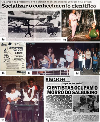
Figure 2. Electron microscopic images of plant cells (a, c) that served as templates for constructing a giant cell model exhibited at the Life Museum, at the Oswaldo Cruz Foundation, Rio de Janeiro (b, d). Images from a transmission electron microscope (a) or scanning electron microscope (c) may be useful to construct 3D models. Cell wall (w), nucleus (n), chloroplasts (c), vacuole (v), mitochondria (m), and Golgi (g).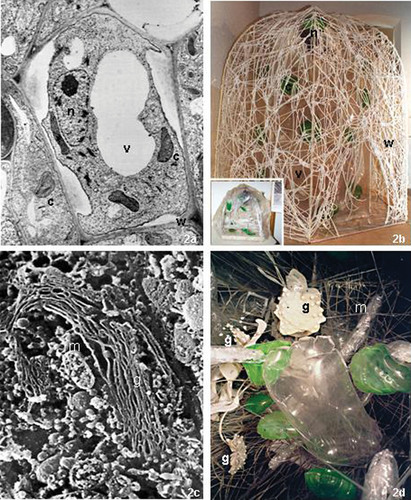
Figure 3. Workshops with teachers for interactive microscope activities (a) and cell modeling (b), and the front cover of the first four modules (c-f) of the series of handout sheets for science activities, “With Science at School.”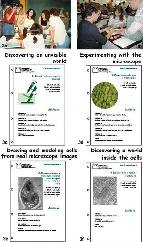
Figure 4. Prototype of games developed with images from light (a) and electron (c) microscopy. In the cell puzzle the microscopy field that can be observed in a real instrument (b) can be reconstructed in the game, where the individual pieces were cut at the cell wall boundaries. In the game about spermatozoid transformation (c) the challenge is to follow the steps in the process of cell differentiation.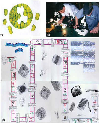
DEVELOPMENT OF HIGH QUALITY IMAGE GUIDE: “COM CIÊNCIA NA ESCOLA”
In all the workshops, printed images were frequently requested by the attending public, to assure full comprehension of what they were seeing through the microscopes. We then rebuilt our original gallery of cell images, made them available in good print quality for the teachers, and prepared a new guidebook for practical activities, Com Ciência na Escola [With Science at School] (Figure 3c-f). In Portuguese, the expression Com Ciência sounds similar to the word consciência, which means“ awareness” or “consciousness.”) The guidebook organized suggestions on laboratory activities using microscopes and changed the “cookbook” view of the traditional guidebooks. In these activities, we offered suggestions on things to do, things to think about, and questions to answer but never provided the answers to the proposed questions, thus creating a real space for teacher creativity and experimentation conducted with their students. Images obtained under conditions similar to those proposed in the protocols were also provided, allowing the students to compare their observations with those of other authors. In addition, some basic procedures of the scientific method were incorporated in all the activities: construct protocol notebooks (Figures 3b and 5a), record the goals, ask questions or pose hypotheses, describe materials and methods used in the experiments, comment on the results obtained, and record the conclusions—which often leads to new questions and new experiments. Students were gradually introduced to data analysis tools and information about different equipment, methods, drawings, or models. Students considered the choice of different experimental models to address questions and to generate data; one of the most frequently used was Elodea (Figure 5b) (Carstensen et al., 1990; Walker, 1994), as well as two more common research organisms, yeast (Manney and Manney, 1992) and C. elegans (Hodkin et al., 1998; Morgan, 1999). Magnetic bacteria or nonpathogenic protozoa can also be used in the classrooms to advance biology education. These organisms are less costly and affected by fewer ethical constraints. The practice of using live experimental model organisms in classroom activities, analyzing results, and discussing conclusions could help to bridge the fields of science and education, simulating the actual process of science advance.
We wanted to learn from elementary- or high-school teachers what questions would improve cell biology in the classroom. We narrowed this down to four questions for the first four modules of the series: How do microscopes work? How can we experiment (and not just demonstrate) using a microscope in the classroom? What do cells look like under an optical or electron microscope? and How are the intracellular structures seen in images obtained using different techniques?
The first modules of the Com Ciência na Escola series start with these four questions (examples of front covers in Figure 3, c-f). The modules were initially available on CD-ROM as files to print, since common PCs and printers are available to the teachers in almost every school, at least in the major cities in Brazil. The printed versions are a series of individual handouts, each one proposing three to five different practical activities on a common subject that can be selected for use by the teachers or the students. In some of the modules, light and electron microscopic images are proposed as templates for different activities such as (a) outlining all the inner structures of plant and animal cells, protozoa, and bacteria (Figure 6); (b) sculpting inner structures with modeling clay in two or three dimensions, maintaining the natural scales of the real images (Figure 7, a-c); (c) modeling cells with balloons or condoms filled with water to let the students “feel” what the consistency of a cell should be (Figure 7, d and e); (d) modeling inner cell organelles to scale, to construct 3D models of cells (Figure 2, b and d); and (e) measuring cells and intracellular components (Figure 5, c-e).
According to our pilot test of these activities, none of 30 teachers had ever drawn his or her own sketch of a cell based on a real microscope image (Figure 6, a and b). By overlaying transparency sheets and using color pens, the teachers clearly highlighted the complexity of cytoplasm and nuclear contents in the real images. The simplicity of the schematic diagrams that emerged from their drawings were useful to help discover the main different cell compartments, such as the Golgi apparatus, mitochondria, the endoplasmic reticulum, and a diversity of granules. The teachers could see and draw different vesicles and particles and were then confronted with the difficulty of identifing structures without a marker of their function. So a vesicle's identity and function become a problem-solving method of posing new questions instead of an answer (Chiappetta, 1997; DebBurman, 2002). This was an important and integral part of the activities, since both the teachers and the students started posing new questions after answering the initial question. The answering and asking of questions are one of main elements of the scientific thinking and an important skill to develop during science education courses. Students show the same ease (and fascination) as their teachers when tracing the intracellular outlines using microscopic images (Figure 6, c-f). In the example shown in Figure 6, c and d, the difference in size of mitochondria and chloroplast, which are often represented as similar-sized structures in books, came out clearly in the drawing by a 12-year-old student. In the diagram of a bacteriophage drawn by another student (Figure 6, e and f), the association of virus particles and bacterial nucleic material and the great difference in their sizes became obvious. The same images used to generate 2D diagrams allowed a more tactile experience when inner cell compartments were sculpted with modeling clay (Figure 7, a-c) and the students were confronted with all the details and relative sizes and forms of the different cell organelles and compartments. The advantage of these graphical exercises is the emotional involvement of an amusing activity commonly performed by children and recently rediscovered as a pedagogic strategy even for illustrating literature books (Xavier, 1997). Membrane-bound structures and their contents can be easily represented and the relative proportion of each cell compartment can be estimated. Indeed, exercises can be developed with microscopic images, such as measurement of the microscopic field (Ekstrom, 2000, 2001) to estimate the cell size. Although image-processing digital photography software can refine these practices (Ekstrom, 2002), a nondigital and affordable alternative version of such morphometric studies can be performed by weighting the compartment profiles that are drawn and cut (in the example in Figure 5, a-c, the profiles of mitochondria) and relating them to the whole weight of the section studied.
Figure 5. A sheet for the protocol notebook (a) and Elodea as one of the experimental models (b) suggested in the education material. (c-e) An activity to estimate the relative size of the mitochondrial compartment in an animal cell by drawing their contours on a transparency sheet, cutting them out, and weighting the profiles on a balance.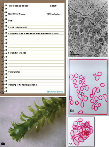
Figure 6. Images versus drawings. Original transmission electron microscopic images of a lymphocyte (a), a plant cell (c), and a bacteriophage with virus (e) and the corresponding 2D diagrams (b, d, and f) prepared by students over the image templates on a transparency sheet.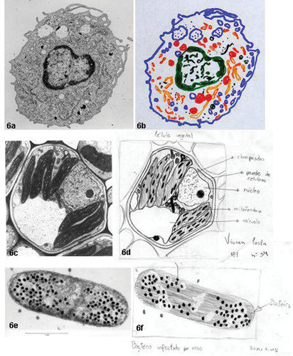
The building of water-filled models (Figure 7e) was always a wonder for both teachers and students. For these models, the relative scales and proportions of inner cell compartments are not the focus; instead, the important novelty is the mobility of different globules and granules that can be represented by balloons of different sizes and colors filled with water. Filling the whole “cell” with water, with liquids of different densities, or with gels allowed the strange experience of feeling what would be the consistency of a cell if one could touch it. A neuron was modeled and empty plastic pencils were used to simulate the cytoskeleton and sculpt the dendrites (Figure 7, d and e). Implantation cone and axons with nodes of Ranvier were also chosen by teachers to be represented in the model. Watery cell models are educational 3D toys that can be constructed easily to permit different perceptions of the moving/plastic aspect of the cells.
In conclusion, the educational materials reported here incorporate important tools of the scientific research world: the protocol notebook; the use of instruments, equipment, models, and drawings; and the use of common experimental model organisms.
NEW CHALLENGES AND CONCLUSIONS
It has been recognized that the traditional lecture is frequently a passive experience for students and that approaches that enhance their active participation in the learning process can deepen their understanding (Bonwell and Eison, 1991). This was our main goal while preparing our materials. As can be judged by the comments of both teachers and students at the end of the activities, our goals were achieved. Sometimes it was difficult to end the classes when it was time to do so.
Two important new projects evolved from these materials and activities. The first stemmed from the need to construct a digital library of high-quality cell images. This is the goal of the Biodigital project (Araújo-Jorge et al., 2003), which is now partially developed, although it will always be under construction. The second arose from the need for more frequent and profound courses for training science teachers. A program for “Science Education on Biology and Health” (Grynszpan and Araujo-Jorge, 2000), addressing the need for teacher training and the new course, has spawned M.Sc. and Ph.D. courses for science and biology teachers who want to innovate in their own classrooms by interacting with scientists in research centers.
Teaching and research can be mutually beneficial rather than competitive pursuits (Huang, 2000). Indeed, in Brazil, most scientific research is conducted at public universities (Leta et al., 1997) and, therefore, by researchers who are also teachers. The interest in how to integrate research and education is stronger than ever and is spreading all over the world, due to greater public awareness of the importance of science to economic development and social well-being. The U.S. National Science Foundation (1996) has initiated a number of programs with the explicit purpose of promoting efforts toward such integration. More significantly, the NSF has implemented a new policy to focus the merit review criteria for research proposals into two categories: (a) intellectual merit—the questions for this criterion include,“ What is the likelihood that the project will significantly advance the knowledge base within and/or across different fields?” and (b) broader impact, to include education as a desired research activity. Here, the emphasis on education is shown in questions such as, “How well does the activity advance discovery and understanding while concurrently promoting teaching, training, and learning?” and “Does the activity enhance scientific and technological literacy?”
Huang (2000) emphasized the impact of an innovative curriculum on academics:
On many campuses, protocols to encourage faculty members to engage more seriously in formal teaching have been established. At the Johns Hopkins University, for instance, a teaching portfolio is suggested for those who come up for promotion. A typical teaching portfolio might consist of the following items: A statement of your teaching philosophy; educational programs you have developed; curricular innovations; teaching material developed; student evaluation; published scholarly education activity. This situation is a clear change from the traditional assignment for faculty members to simply offer courses in the form of lecture and/or laboratory exercises. If this reflects a new movement, faculty members are given the signal to be more conscientious about education than ever before on US campuses.
Distance learning and consortium labs are already a reality for research projects. The idea of networking many NMR studies with a base unit, a“ collaboratory,” is an example (Kouzes et al., 1996; Myers et al., 1997). The arrangement, through collaborative demonstrations, promises that one can do experiments and teach at a distance, sharing facilities and talents. Linking nodes of databases and processing and integrating data may be another avenue basic to biology, in research and in education. Attempts to form teaching consortia are under consideration and amendable with the availability of network devices (Kouzes et al., 1996). The interdisciplinary approach to education may, in the near-future, collect scientists into one “virtual room,” a single virtual laboratory. The daily activities of science, such as talking to colleagues and students, reading journals and working in the laboratory, will be conducted in a seamless digital theater.
Figure 7. Image of cells (a, d) serving as templates for sculpting organelles of modeling clay to scale (a-c) or for modeling with toy balloons filled with water (e).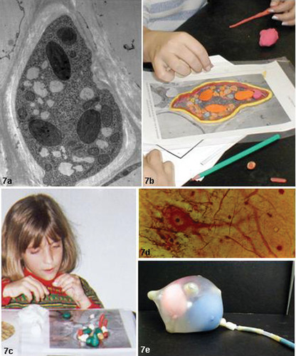
However, good research is doubtful without a good sense of value and sensibility. Cell biology, together with genetics and molecular biology, is vulnerable to challenges in ethical or legal terms for its discoveries and practices. The need to develop a sense of balance and ethical curiosity is part of the education. We must not let the technological revolution drive us to training technicians rather than educating scientists and citizens.
FOOTNOTES
Monitoring Editor: Mary Lee Ledbetter
ACKNOWLEDGMENTS
This work is dedicated to all our colleagues at the Espaço Ciência Viva, where all this begans. It is also dedicated to the memory of two of our masters from the Carlos Chagas Filho Institute of Biophysics, Dr. Hertha Meyer and Dr. Raul Dodsworth Machado, who always supported our open-air outreach education activities with ideas, images, and microscopes. The authors acknowledge A. Malcolm Campbell, Davidson College, North Carolina, for his excellent revision of the English. This work was supported by the Oswaldo Cruz Foundation (Fiocruz) and Conselho Nacional de Desenvolvimento Científico e Tecnológico (CNPq). Cláudia Coutinho and Mauricio Luz are associate professors at the Laboratory of Cell Biology within the Fiocruz agreements with the Universidade Federal Fluminense (UFF) and Universidade Federal do Rio de Janeiro (UFRJ).


