Bringing Developmental Biology to Life on the Web
INTRODUCTION
The process by which a single-celled egg develops into a complex, multi-billion–celled individual has perplexed philosophers and scientists for millennia. Aristotle described chick development in the egg as early as 340 B.C. In the 17th century, Hartsoeker (1694) discovered “animalcules” in human and animal sperm and suggested that this preformed homunculus grew into a child when placed in a woman's womb (Figure 1). Because of the research tools available, developmental biology remained a descriptive science until the second half of the twentieth century. Since then, new techniques adapted from cell biology, molecular biology, and genetics have enabled researchers to make great strides toward elucidating one of the greatest mysteries of life.

Figure 1. Hartsoeker's homunculus shows a preformed human contained in a sperm (Hartsoeker, 1694). From http://en.wikipedia.org/wiki/Image:HomunculusLarge.png/.
The multimedia capabilities of the Web provide an ideal medium for bringing to life for students the dynamic molecular and morphological processes of developmental biology. Movies and animations can assist learners in visualizing these complex, time-dependent processes, augmenting the static illustrations in texts. This Feature describes sites that students can use to supplement and extend what is taught in class as well as materials that instructors can incorporate into lecture or laboratory, with special emphasis on introductory-level biology and developmental biology courses.
FLYING THROUGH DEVELOPMENT
Of all the websites I explored, the FlyMove site stands out as the one that best utilizes the Web to support learning about multiple aspects of development (http://flymove.uni-muenster.de/; Figure 2). This well-designed site, produced by a team at the Institut für Neurobiologie at the University of Müenster (Müenster, Germany), is divided into five sections. 1) The Stages section covers the 17 embryonic stages of Drosophila development. The “Embryogenesis, in vivo” time-lapse movie, which is accessed by a link on the first page of this section of the site, shows fly development through all of the stages. The movie could be used with younger students to help convey the dynamic cell movements that take place during embryogenesis. The video would benefit from the addition of narration because it is difficult to read the text about each stage while watching it, though viewers can pause the movie to read. 2) In Processes, one can learn about pattern formation, morphogenetic movements, and cellular processes. These are the only online materials I found that addressed such topics in an accessible manner for students. 3) The formation of the major organ systems is addressed in the Organogenesis section. 4) Although much of the Genetics section is still under construction, it currently includes information on how to handle flies and perform crosses. I look forward to seeing the content that will be included in sections on mutations, Mendel's Laws, mapping, transposons, genetic screens, and genetic circuits. 5) The Methods section also is being built, but so far includes two important laboratory techniques: cell transplantation, which is a tool for studying cell fate determination, and DiI labeling, which enables researchers to trace the embryonic development of individual progenitor cells as well as study the morphology of their fully differentiated progeny. In each section, animations, movies, line drawings, and photographs are accompanied by explanatory text. Although those who work with Drosophila will be familiar with embryo size at each stage, the inclusion of scale bars in visual materials would be useful for students. At the bottom of each page a Genes Discussed section summarizes information about the genes mentioned, along with links to additional information. Unfortunately, none of the ones I tried worked.
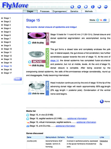
Figure 2. The FlyMove website addresses the molecular and morphological processes of development.
Several student support materials are incorporated into the website. The homepage for the Tour section includes an animated tour of the website, which introduces the user to the navigation and available features. I wish more content-rich sites had a similar feature. Two subsections of the Tour provide resources that could be used at the beginning of fruit fly labs. The History subsection provides a brief, animated introduction to the advantages of using Drosophila as a model organism and to its developmental stages and life cycle. The Life Cycle subsection includes an animation that begins “If you are under 18—please leave the slide show, otherwise click inside the window to see more.” This tongue-in-cheek warning should definitely pique the interest of high school students. A Glossary serves as an online reference, providing brief definitions, some with diagrams. Quizzes in several sections of the site offer opportunities for students to test their knowledge. Instructors will find the searchable database in the Media section a helpful resource for finding and downloading any of the visual materials on the site.
WHERE DID I COME FROM?
From an early age, children are interested in how they came to be and in their own body's development. This interest provides an excellent “hook” for engaging students in biology during middle and high school, as well as beyond. When looking at the websites described below, it is helpful to know that Carnegie stages (numbered 1–23) are used by embryologists to describe the developmental maturity of human embryos, based on their external features.
The Multi-Dimensional Human Embryo site was developed by Bradley Smith at the University of Michigan (Ann Arbor, MI; http://embryo.soad.umich.edu/; Figure 3). It has beautiful, rotating, 3D images of Carnegie stages 13–23 (approximately 28–56 days after ovulation), produced using magnetic resonance microscopy. Organs such as heart, liver, or lungs are highlighted in individual movies, allowing one to focus on a particular organ. The black-and-white QuickTime movies (Apple Computer, Cupertino, CA) are complemented by color photographs made with a low-power microscope. A scale mark on one of these for each stage provides information on actual size. In addition, those interested in viewing coronal, sagittal, and axial sections can do so by clicking on the MRI Slice Selector images. A brief description of each stage provides the embryo's postovulatory age, length measurement, and distinguishing developmental criteria. A pseudo time-lapse movie, linked on each page, shows development through all of the stages and would be appropriate to use even with younger students.
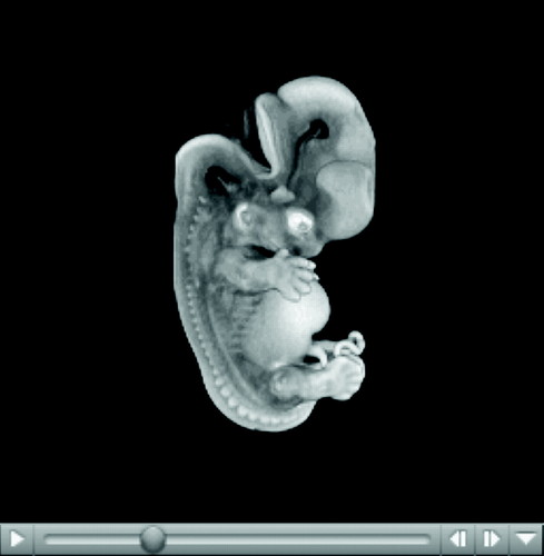
Figure 3. A 3D image from the Multi-Dimensional Human Embryo website.
The Visible Embryo was the best site I found for introductory-level students and the public on human development from conception to birth (http://www.visembryo.com/; Figure 4). Although it does not include animations or movies, it does employ several interactive interfaces that facilitate navigation. Clicking on Pregnancy Timeline in the menu bar at the top of the page accesses a week-by-week description of development. Scrolling over the number for each of the 40 weeks of pregnancy provides one developmental milestone for that stage. Clicking on the number takes you to a page that briefly describes in nontechnical language the developmental processes taking place at that time in different parts of the body, such as the head and neck, abdomen, limbs, pelvis, thorax, and skin. The site says that it includes movies but I was unable to find them. However, the movements of cells and tissues that take place during development are noted to some extent. Each page also includes an image of the developing embryo, fetus, or infant as well as information about its size. Although these images include both metric and English rulers, close examination reveals that the images often are not the correct size with respect to the rulers. However, they do provide an indication of relative size and growth between weeks.
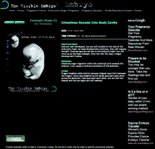
Figure 4. The Visible Embryo website provides a week-by-week summary of human development.
This site's navigation is challenging, because clicking on Home in the top menu bar does not take you back to http://www.visembryo.com/, where the news section is located. The news section is updated daily with synopses of the latest stories about human development and pregnancy, with links to the original articles. Articles from the popular press and scientific journals are separated and an archive is available. This feature should be very useful to instructors, enabling them to easily update their lectures.
Because of the small font and primarily text-based pages, this site is probably most useful for students to explore on their own, rather than for classroom projection. Teachers may want to point out to students that some of the links on the site are to advertisements by Google and so need to be viewed critically as to the quality of the content.
INTO THE LABORATORY
Although animations and videos can elucidate the complex processes of development, it is important that students also have the opportunity for laboratory experiences. I still remember my sense of awe the first time I watched sea urchin (Echinoderm) eggs being fertilized and saw the fertilization membranes arise. It also was fascinating to observe the embryos develop to the pluteus stage.
Stanford University (Palo Alto, CA) provides a multimedia approach to laboratory explorations utilizing Echinoderms. I particularly liked how the Sea Urchin Embryology site (http://stanford.edu/group/Urchin/; Figure 5) provides guidance for several levels of inquiry, from observation-based through guided and full-inquiry experiments. Step-by-step instructions guide students as they learn basic procedures for working with the system. Linked animations show techniques and what is happening at each step. The animations are very simple graphically, but do convey their intended information. Time-lapse movies of several stages of development are included too. The animations and videos that are linked from the labs also are available in a developmental timeline called the Path of Development. I appreciated that this section of the website is available in Spanish, facilitating use in the growing number of classrooms with English language learners. Questions at the end of each lab require students to think about development and what they have observed. The techniques and observations in the basic labs can provide students with the skills they will need to continue with higher levels of inquiry. Extensive resources for instructors are provided to facilitate implementation of the labs. Although this website was developed for the high school level, it also would be useful to college faculty and laboratory coordinators.
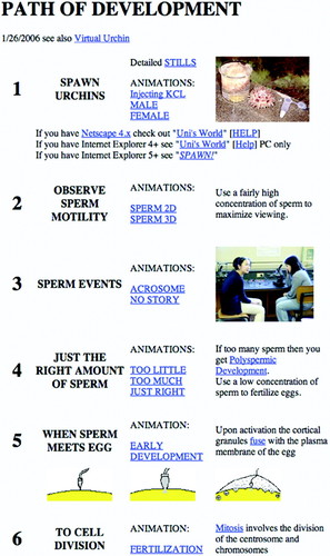
Figure 5. The Sea Urchin Embryology website includes labs with several levels of inquiry. The Path of Development assists students in understanding each stage.
The Virtual Urchin site, also from Stanford, is in the pilot stage (http://virtualurchin.stanford.edu). When completed, it will provide a nice complement to the laboratory site. Sections include Urchin Anatomy, Sea Urchin Fertilization and Development, Predator-Prey, and Microscope Measurement. The later interactive virtual lab could be used as a complement to any lab in which students are learning to measure objects using a compound microscope.
ADDITIONAL DEVELOPMENTAL BIOLOGY WEBSITES
Welcome to the Dynamics of Development!
Welcome to the Dynamics of Development! (http://worms.zoology.wisc.edu/embryology_main) was designed as a supplement to Jeff Hardin's Introduction to Animal Development course at the University of Wisconsin-Madison. Tutorials for echinoderm and amphibian provide materials for several stages of development with brief descriptions, links to Glossary words, and movies. The index allows the user to locate quickly a topic of interest, or they can click through each section.
C. elegans Movies
C. elegans Movies (http://www.bio.unc.edu/faculty/goldstein/lab/movies.html) has many movies of Caenorhabditis elegans development with varying amounts of explanatory text. A 4D movie allows the user to view development through time with the ability to move up and down through several focal planes.
All I Really Needed To Know I Learned during Gastrulation
All I Really Needed To Know I Learned during Gastrulation (http://www.sdbonline.org/fly/gilbert/gilbert01.htm) provides a distillation of key concepts about development, with humorous images. The PowerPoint lecture (Microsoft, Redmond, CA) was presented by Scott F. Gilbert (Swarthmore College, Swarthmore, PA) at the 64th Annual Meeting of the Society for Developmental Biology in San Francisco, CA, on July 29, 2005.
ACKNOWLEDGMENTS
I thank A. Malcom Campbell for critical comments and helpful suggestions on this article.


