WWW.Cell Biology Education
Cell Biology Education (CBE) calls attention each quarter to several web sites of educational interest to the life science community. CBE does not endorse or guarantee the accuracy of the information at any of the listed sites. If you want to comment on the selections or suggest future inclusions, please send a message to [email protected]. The sites listed below were last accessed on November 10, 2002.
DEFORMED AMPHIBIAN RESEARCH AT HARTWICK COLLEGE
http://info.hartwick.edu/biology/def_frogs/index.html
An excellent and interesting implementation of the scientific method is found at this interactive site created and maintained by Stanley Sessions at Hartwick College in Oneonta, New York. Reports of malformed frogs in North America have been increasing over the last 10 years, with at least nine web information centers tracking the decline in frog populations. One of the centers that tracks information and reports research on the phenomenon is located at Hartwick College. Sessions has created a highly useful web site that presents the amphibian malformation problem in a highly useful way to students and biology educators.
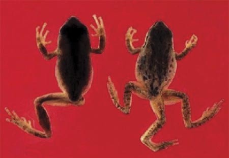
Figure 1. Frogs that demonstrate leg deformities. From the laboratory of Stanley Sessions of Hartwick College. Reprinted by premission of Stanley Sessions (November 10, 2002).
The exercise allows a student to move through the malformation evidence in a directed free-form manner. The student is given some visual evidence of malformations (see Figure 1) and then offered seven hypotheses to explore. Detailed evidence with references is provided for four of the hypotheses. Two explanations developed are the well-known effects of vitamin A and ultraviolet light on vertebrate limb growth. This captivating exercise allows the student to draw a conclusion that most would never consider. The scientific method is beautifully explored in a real-time problem. The deformed amphibian web site provides an excellent entreé into the inquiry approach to laboratory learning. There are also some wonderful examples of photomicroscopy.
C. elegans MOVIES
http://www.bio.unc.edu/faculty/goldstein/lab/movies.html
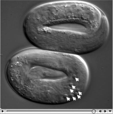
The international group of researchers using Caenorhabditis elegans as a model has just announced the complete genome sequence, consisting of just over 100 million base pairs, for the worm. Supporting this effort, Robert Goldstein at the University of North Carolina at Chapel Hill has assembled thirty-five movies demonstrating various activities of C. elegans. Drawn from 20 laboratories worldwide, the movies provide the developmental sequences of normal and mutant worms (see Figure 2). Topics include visualizations of cytoplasmic flow, P granules, tubulins, and histone movement. Techniques using POP-1, PIE-1, and PES-1 reveal highly informative developmental events. The worms are shown crawling, rolling, mating, ovulating, trafficking, and in seizure. From spermiogenesis to calcium transients, the Goldstein collection provides a wonderful resource of C. elegans movies. Even three RNA interference (RNAi) screens are referenced. For an instructor who wishes to explain to students why a roundworm was the focus of a 2002 Nobel prize, this web site is a good place to begin.
Figure 2. Developing C. elegans showing apoptosis at arrowheads. From the laboratory of Robert Goldstein of the University of North Carolina at Chapel Hill. Printed by permission of Robert Goldstein (November 10, 2002).
MICROBE LIBRARY
http://www.microbelibrary.org/
This source of educational information is part of the Microbiology Education Library maintained by the American Society for Microbiology. The information is organized into four areas: visual resources, curriculum resources, reviews, and articles. As of September 2002, 324 visual images were associated with 223 entries, all with legends and annotations. There are currently 43 classroom and laboratory entries available. A search engine is provided that makes use of headings such as topics, groups of organisms, core concepts, and pedagogy keywords.
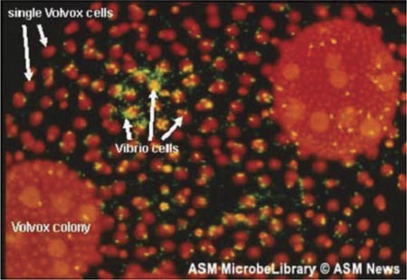
Figure 3. Vibrio cholerae bacteria among Volvox. From Rita Colwell of the National Science Foundation. Printed by permission of the American Society of Microbiology (ASM) and ASM News (November 11, 2002).
Figure 3, from Rita Colwell, is an example of the high-information content graphics available from the resource. Animations and videos accompany the library of still images. One of the curriculum resources is an exercise titled “Do-It-Yourself Immunoglobulin Gene Rearrangement.” It is a highly effective, low-cost, and simple demonstration of gene rearrangement, a perfect way to introduce the subject to a class. Another curriculum resource is titled “Virology, Genome Sequencing, and Bioinformatics.” The various parts and pieces of this laboratory exercise can be downloaded from the site. There is a wonderful review article that could be titled “Who Killed C. elegans?” The actual article deals with cyanide production by P. aeruginosa and its effect on worms. The material could be developed into a case study concerning cystic fibrosis. One of the articles available, titled “Significant Events of the Last 125 Years,” is a delightful history of microbiology with links to support materials. This URL is a superb educational resource, and it is growing in content and usefulness.
NATIONAL SCIENCE DIGITAL LIBRARY
http://about.nsdl.org/
The National Science Digital Library (NSDL), funded by the National Science Foundation, will come on-line during early 2003. The NSDL represents one of the most important science education developments of the next 10 years. Currently the site provides a tour of what is to come. You may want to be one of the first to explore this new educational resource.


