WWW.Cell Biology Education
Each quarter, Cell Biology Education calls attention to several Web sites of educational interest to the life science community. The journal does not endorse or guarantee the accuracy of the information at any of the listed sites. The sites listed below were last accessed on October 5, 2003.
THE HISTORY OF MITOSIS
“Mitosis consists of four stages that are named in sequence: prophase, metaphase, anaphase, and telophase. In between mitotic divisions is interphase, the period of time during which DNA is replicated.” The two previous sentences along with their supporting material verge on a litany and a catechism for biology. The column for this issue will focus on Web resources for teaching and learning about mitosis.
Why do we call asexual cell division “mitosis”? Who created the names “prophase,” “metaphase,” “anaphase,” and “telophase?” Sir Henry Harris, a pathologist from the University of Oxford, provides a wonderful accounting of the history of mitosis in his book The Birth of the Cell (Yale University Press, 1999, ISBN 0-300-07384-4). Supplementing Harris' reference are two Web sites using timelines that put these morphological descriptors of cell division in their historical place.
Electronic Scholarly Publishing http://www.esp.org/timeline
Electronic Scholarly Publishing (ESP) is an effort established by Robert J. Robbins. Two of the projects of ESP are a classical collection of genetics papers and a genetics timeline. If you would like to have Mendel's 1866 paper, it is here. ESP's genetics timeline places the discoveries leading to the description of mitosis into chronological perspective. Beginning in 1750, the timeline juxtapositions science with world events. For mitosis history, the timeline between 1875 and 1895 is especially interesting.
Lasker Foundation http://www.laskerfoundation.org/news/gnn/timeline/timelinetop.html
The Lasker Foundation also has a nice timeline for genetics and genomics. Not as ranging as the ESP timeline, the Lasker timeline provides background information on some of the individuals including Walther Flemming, the person who coined the term “mitosis” in 1882. The Lasker timeline can be downloaded as a 211-page PDF file and includes a nice biography and picture of the first person to recognize that chromosomes undergo longitudinal division during mitosis, that is, Walther Flemming.
The various origins of the terms associated with mitosis are listed in these timelines. W. Schleicher developed the term “karyokinesis” in 1878. Eduard Strasburger created the terms “prophase,”“ metaphase,” and “anaphase” in a paper published in 1884. Heinrich Wilhelm Gottfried Waldeyer gave us the term“ chromosome” in 1888. Surprisingly the ending phase of mitosis,“ telophase,” was introduced by Martin Heidenhain in 1894. It is curious that 10 years lapsed between prophase and telophase in terms of naming. And finally an H. Lundegardh put “interphase” into the mitotic lexicon in 1913. This history of cytology can be found in greater depth in the Harris text and in the 1911 Encyclopedia Project.
The 1911 Encyclopedia Project http://93.1911encyclopedia.org/C/CY/CYTOLOGY.htm
This 1911 Encyclopedia article is interesting as a historical document dealing with what was then the recent history of the development of cytology. The text of the whole 1911 Encyclopedia is available at the above Web site. Of a related nature, if you have an interest in structures that are named after famous scientists, then you might what to check a medical eponym site.
Dictionary of Medical Eponyms http://www.whonamedit.com/
Almost 6500 medical eponyms are listed at this URL. This site is mentioned because a nice biography of Waldeyer, the primary source for the term“ chromosome,” may be found here.
TEACHING MITOSIS TODAY
A more recent history of mitosis research may be found in a Nature article by T.J. Mitchison and E.D. Salmon. (2001. Mitosis: a history of division. Nature Cell Biology 3(1): E17–21.) A PDF version of this article may be found at the following URL.
T.J. Mitchison Lab Page http://mitchison.med.harvard.edu/Publications.htm
It was this paper in Nature Cell Biology that inspired this column. The report provides an excellent pictorial view of the evolution of thought concerning how the mitotic spindle is formed and generates force. This five-page paper will bring you to the edge of contemporary ideas on mitosis.
John Kimble's Electronic Biology Textbook http://users.rcn.com/jkimball.ma.ultranet/BiologyPages/M/Mitosis.html
A good place to start our tour of Internet mitosis sites is Kimble's textbook description of mitosis. There are many hypertext links to other locations that provide definitions and examples for key terms encountered in the vocabulary of mitosis.
BioWeb at University of North Carolina at Charlotte http://www.bioWeb.uncc.edu/biol1110/Stages.htm
Biology 1110 at UNCC is the lab course that supports nonmajors biology. The course Web site has an excellent traditional exercise on mitosis supported by images such as found in Figure 1.
VIEWING MITOSIS
A search of the Internet can provide excellent illustrations and photographs of mitosis that move beyond the traditional text and lab materials. Below is a brief sampling of some of these wonderful and often esthetic views of cell division.
Biology 304 Homepage http://www.micro.utexas.edu/courses/levin/bio304/genetics/celldiv.html
This Web site supports a majors-based course at the University of Texas at Austin titled Biology 304—Ecology and Evolutionary Biology. The first image on this Web page has a traditional view of plant mitosis and that figure is reproduced here as Figure 2.
Biology Teaching Organisation http://www.bto.ed.ac.uk/courses/year1/mac1h/gallery/index.html
This site is located at the University of Edinburgh. The image gallery supports many courses including a first-year course in Molecules and Cells. Figure 3 is an example of a beautiful mitotic figure fluorescently stained and found at the Edinburgh site.
Figure 1. A standard depiction of whitefish and onion mitosis. The chromosomes of anaphase are clearly realized in each. Courtesy of Dr. Mark Clemens of the Department of Biology, University of North Carolina at Charlotte.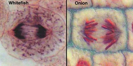
Figure 2. The four stages of plant mitosis are seen in this composite with the chromosomes staining blue and the spindles red. Courtesy of Dr. Andrew Bajer, Professor Emeritus of the University of Oregon.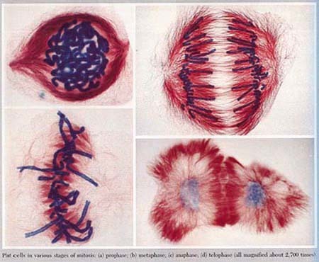
The Vivek Malhotra Lab http://www-biology.ucsd.edu/labs/malhotra/SciencePictures.htm
The Web site listed here has been developed by Dr. Vivek Malhotra's Lab in the Division of Biological Sciences at the University of California, San Diego. There are images at this site that answer a question which students often ask: what happens to the cell organelles during mitosis. Figure 4 reveals Golgi being partitioned during mitosis.
Figure 3. An early anaphase with red stained chromosomes and green spindle microtubules of a mammalian kidney cell is illustrated. Courtesy of Professor William C. Earnshaw, Institute of Cell and Molecular Biology, University of Edinburgh.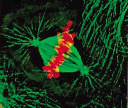
Figure 4. This photomicrograph appeared on the cover of the April 17, 2000 issue of the Journal of Cell Biology. The caption states: “Laser scanning confocal fluorescence image showing the organization of the Golgi membranes during various stages of the cell cycle. Normal rat kidney cells (courtesy of Dr. Lippincott-Schwartz) expressing galactosyl transferase-YFP (shown green) were costained with the DNA-specific stain propidium iodide (shown blue) and antitubulin antibody (shown red). In the nondividing cells the Golgi membranes are found in the perinuclear area.” The article associated with the cover image may be found at http://www.jcb.org/cgi/content/short/149/2/331. Courtesy of Vivek Malhotra of UCSD and the Rockefeller University Press.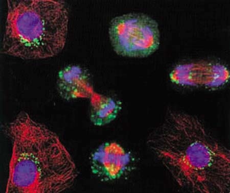
John Runions' Home Page http://www.plantsci.cam.ac.uk/Haseloff/JohnRunions/Web/Lineage.html http://www.plantsci.cam.ac.uk/Haseloff/JohnRunions
John Runions was a post-doctoral fellow in the lab of Jim Haseloff at the University of Cambridge when the images found at the URL above were made. Accompanying the beautiful pictures of Arabidopsis root, Runions' site has explanations of the techniques employed to produce the wonderful pictures. The images here help a student understand the lineage of cells that are rapidly dividing in succession as illustrated in Figure 5.
Control of Cell Division, Course BS313 http://www.teaching-biomed.man.ac.uk/ramsay/Overv.htm
The School of Biological Sciences at the University of Manchester offers a course titled Control of Cell Division. The course has a supporting Web site that offers information about the control of mitosis at nine different time points. By clicking on one of the time points arrayed in a diagram, clear organizational illustrations appear that indicate the relationships of such compounds as cdc2, cyclin B, wee1, and cdc25 in the molecular control of mitosis.
Figure 5. Cell lineage analysis of Arabidopsis root apical meristem is demonstrated here. Yellow fluorescent protein is bound to histone 2B and reveals all the cell nuclei that are related to one another. Courtesy of Dr. C. John Runions of the Department of Biological and Molecular Sciences at Oxford Brooks University.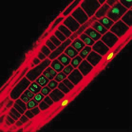
Cande Lab http://mcb.berkeley.edu/labs/cande/mitosis.html
Dr. W. Zacheus Cande is a member of the Department of Molecular and Cell Biology at the University of California at Berkeley. His lab site has some wonderful images of mitosis including rotating three-dimensional reconstructions of mitotic chromosomes and represented by Figure 6.
Cyr Lab http://www.bio.psu.edu/People/Faculty/Cyr/Lab/coolmovies/coolmovies.htm
The lab of Dr. Richard J. Cyr of the Department of Biology, Penn State University, explores questions concerning the cellular basis of plant morphogenesis. At the URL above are a number of movies that show Golgi movement, cycle cell movement, and mitosis. The movies found there are in a variety of Web formats. Figure 7 is representative of the imagery found at this site.
Cytographics http://www.cytographics.com/gallery/gal.html http://www.cytographics.com/gallery/clips/prophase.mpg http://www.cytographics.com/gallery/clips/Celldiv.mov http://www.cytographics.com/gallery/clips/Sickcell.mov
Dr. Jeremy Pickett-Heaps has been producing awe-inspiring views of cells for years. He and his colleagues have formed a distribution company of educational materials called Cytographics. The first URL takes one to a gallery of video resources. The last three sites listed above link to direct downloads of short videoclips of cells in division; the clips may download slowly. The fourth site provides a view of misdivision in mitosis and shows a chromosome going astray during division. Figure 8 is typical of the quality of the images found at the site.
Figure 6. This still image was taken from a set of five rotating movies of yeast cell chromosomes in division as seen from reconstructed laser confocal microscopy data. Courtesy of Zacheus Cande of the University of California, Berkeley.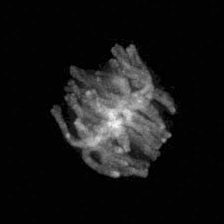
Figure 7. “The image is of a tobacco cell (BY-2) that has been stably transformed with a Golgi marker in the red channel (nactylglucosamine:dsRed) and a microtubule marker in the green (microtubule binding domain of MAP4:GFP). Plant cells have a barrel shaped anastral spindle and so it looks a bit different (from) animals.” The movie represented by this anaphase figure will form a phragmoplast, which plant cells use in cytokinesis to lay down a new plate. Description and figure courtesy of Dr. Richard Cyr, Department of Biology, Pennsylvania State University.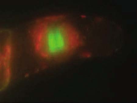
Figure 8. A still image from the videoclip of newt lung cells in division. The chromosomes are on the metaphase plate. Courtesy of Julianne Pickett-Heaps of Cytographics.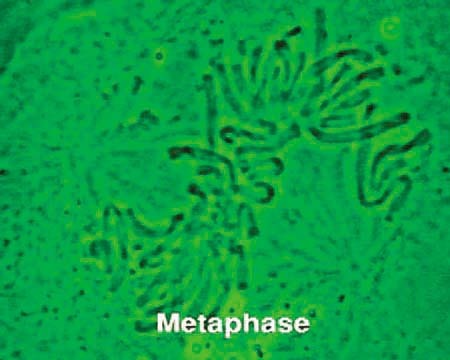
MBC Online Sampler http://www.molbiolcell.org/misc/video1.shtml
The American Society for Cell Biology publication Molecular Biology of the Cell has an online sampler page of videos. One of the videos in the collection represents mitosis in newt lung cell using a new type of microscope. The video represents the work of Shinya Inoue and Rudolf Oldenbourg and the paper supporting Figure 9 seen below appeared in Molecular Biology of the Cell 9(7): 1603–07, July 1998. The text of the article may be found at < http://www.molbiolcell.org/cgi/content/full/9/7/1603>. The video images are of exceptional quality.
Figure 9. The figure caption from the article above states: “Mitosis in tissue-cultured lung cell of a newt, Taricha granulosa, recorded with the new Pol-Scope.” Courtesy of the American Society for Cell Biology.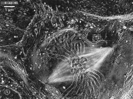
The Kevin Sullivan Lab http://www.scripps.edu/cb/sullivan/movies/ks701.mov http://pingu.salk.edu/~wahl/ks701.mov
Figure 10. This still image comes from a 3-D reconstruction and rotation where the centromeres of a living human cell can be seen during interphase. Courtesy of Kevin Sullivan of Scripps Research Institute.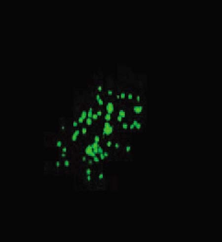
Dr. Kevin Sullivan of the Department of Biology at Scripps Research Institute, working in conjunction with the lab of Geoffrey Wahl at the Salk Institute, has developed a technique to follow the centromeres during Interphase. Using GFP probes and centromere binding protein, the centromere satellite DNA can be visualized as in Figure 10 and provide insight into the activities of the cell before the mitotic process begins.
Mitosis World: the Lab of Ted Salmon http://www.bio.unc.edu/faculty/salmon/lab/mitosis/prometspinfixed.mov http://www.bio.unc.edu/faculty/salmon/lab/mitosis/mitosis.html
Dr. Edward D. Salmon of the Department of Biology at the University of North Carolina at Chapel Hill and his lab work on chromosome movement. They have created a meta-site for mitosis. The movie section of the site named Mitosis World has ten excellent movies depicting different aspects of mitotic division and an image from one of the movies may be seen in Figure 11.
Figure 11. A Differential Interference Contrast Microscopy still image was taken from a 43-min time-lapse movie of newt lung cell mitosis. Courtesy of Ted Salmon of the University of North Carolina at Chapel Hill.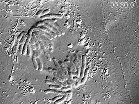
PEER INSTRUCTIONAL MATERIALS
Two student mitosis Web sites were especially interesting and are found below.
Troy High School http://www.troy.k12.ny.us/thsbiology/skinny/skinnymitosis.html
This site is maintained by Troy High School in Troy, New York. The image seen in Figure 12 is taken from an attractive and understandable animation of mitosis. This high school also has online labs to perform onion root tip preparations.
Figure 12. The pink chromosomes move with the green spindle apparatus. Courtesy of Joseph Dibari, a Biology Instructor at Troy High School, Troy, New York.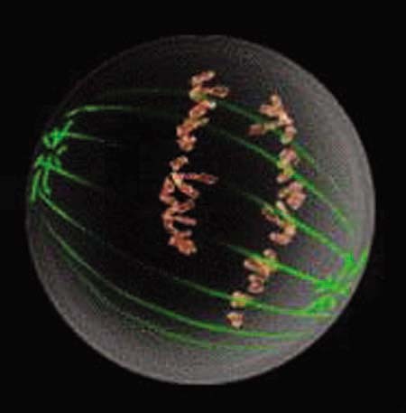
Haverford College Biology 300 http://www.haverford.edu/biology/Courses/bio300/BioGallery02/presentations/CrozierJohnson.htm
Figure 13. Jurkat cells to which DNA has been stained with DAPI. Three stages of mitosis are seen. Met = metaphase; Ana = anaphase; and Telo = Telophase. Images courtesy of Dr. Karl Johnson, Department of Biology, Haverford College.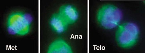
Haverford College biology juniors work on special research topics during one quarter. For their project Kathryn Crozier and Brandon Johnson produced some immunofluorescence images of cells in mitosis and examples may be seen in Figure 13.
It is heartening to consider that undergraduate students can perform such techniques to visualize mitotic events when 120 years ago Flemming, Schleicher, and Strasburger were just trying to name the stages of mitosis.
Thank you for taking this tour through Web resources for studying mitosis. If you want to comment on the selections or suggest future inclusions, please send a message to [email protected].


