WWW.Cell Biology Education
Each quarter, Cell Biology Education calls attention to several Web sites of educational interest to the life science community. The journal does not endorse or guarantee the accuracy of the information at any of the listed sites. The sites listed below were last accessed on December 26, 2003.
CELL SIZE
The interplay of size, growth, division, and diffusion by cells is a common topic in Introductory Biology classes, though often covered superficially. This issue's column will focus on teaching resource materials about cell size. The review begins with a list of four recent articles on cell size that are available as open access PDF files. These articles provide an overview as to the latest thinking about the topic and will be discussed at the end of this article.
C. M. Coelho and S. J. Leevers (2000). Do growth and cell division rates determine cell size in multicellular organisms? J. Cell Sci. 113, 2927-2934. ( http://jcs.biologists.org/cgi/reprint/113/17/2927.pdf)
S. J. Day and P. A. Lawrence (2000). Measuring dimensions: the regulation of size and shape. Development 127, 2977-2987. ( http://dev.biologists.org/cgi/reprint/127/14/2977.pdf)
I. J. Conlon and M. C. Raff (2003). Differences in the way a mammalian cell and yeast cells coordinate cell growth and cell-cycle progression. J. Biol. 2(1): Article 7. ( http://jbiol.com/content/pdf/1475-4924-2-7.pdf)
E. Hafen and H. Stocker (2003). How are the sizes of cells, organs, and bodies controlled? PloS Biol. 1, 319-323. ( http://www.plosbiology.org/plosonline/?request=get-document&doi=10.1371%2Fjournal.pbio.0000086)
GIVING DIMENSION TO THE CELL
The Biology Project http://www.biology.arizona.edu/cell_bio/tutorials/cells/cells2.html
The Biology Project at the University of Arizona is a good place to begin our review of Web resources on cell size. In a brief tutorial on “Size and Biology” (Figure 1), the sizes of animal and plant cells are related to bacteria, virus, and molecules. Information is also provided about light and electron microscopy as they relate to visualizing cells.
Figure 1. Relative sizes of cells. Image courtesy of Dr. William Grimes, Department of Biochemistry, University of Arizona.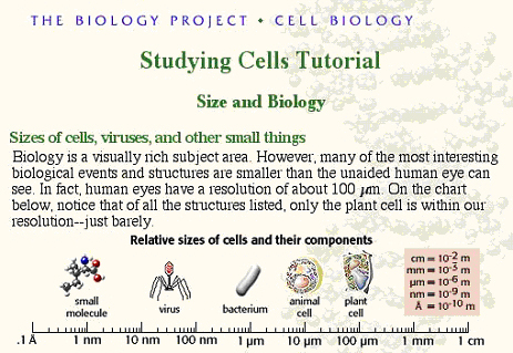
Cells Alive http://www.cellsalive.com/howbig.htm
The Web site Cells Alive has been providing interesting Web-based educational graphics for ten years. Figure 2 provides a representation of the navigation screen for viewing cell dimensions. By clicking on the magnification button, various items from pollen to E. coli are given size context by relating each to the head of a pin. Image courtesy of Jim Sullivan, Quill Graphics, 568 Taylor's Gap Road, Charlottesville, VA 22903.
Biology 1000—Kean University http://www.kean.edu/~biology/cellcalc.html
Dr. Bruce Reid of Kean University has created a simple method for calculating cell size using a ruler and microscope. Figure 3 illustrates how the microscope magnification is determined with a transparent ruler (upper view). Cells are then counted (lower view) and resultant data entered into an interactive calculator that gives the average cell size in millimeters and microns. Image courtesy of Dr. Bruce Reid, Dept. of Biology, Kean University, Union, New Jersey.
GEOMETRY, DIFFUSION, AND CELL SIZE
Many lab activities that explore cell size and shape are based on the geometry of the cell; to be more specific, the relationship between surface area and volume. Working with cell size in this manner is one of the few times that geometry is discussed in introductory biology classes. Three sites are listed below that address geometry issues of cell sizing.
Figure 2. Navigation screen for viewing cell dimensions.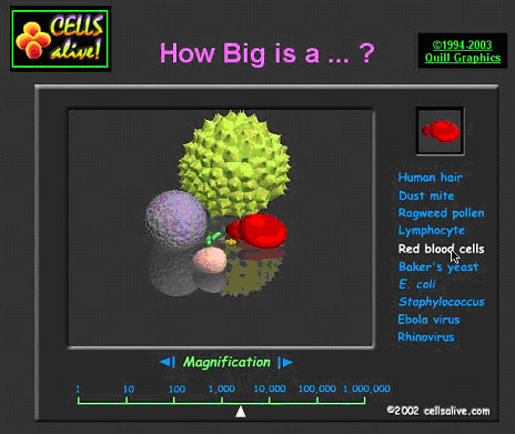
World Builders http://curriculum.calstatela.edu/courses/builders/lessons/less/les4/cellsize.html
Dr. Elizabeth Viau of Cal State Los Angeles asks the question: “How big can cells get?” She provides some graphics that explore cell geometry in a straight-forward lab activity. Three geometric shapes are explored with calculations.
Figure 3. Simple method for calculating cell size. Ruler (top). Cells to be counted (bottom).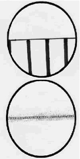
Secondary Classroom Science Concepts http://educ.queensu.ca/~science/main/concept/biol/b02/b02lapl4.htm
The same approach as above is echoed in a lab sizing activity developed at Queens University in Ontario: “Could the world really be overtaken by a giant amoeba?” is the catch phrase used to draw students into the exercise.
Fellows Collection: Access Excellence @ the National Health Museum http://www.accessexcellence.org/AE/AEC/AEF/1996/deaver_cell.html
James Deaver provides a nice hand-on exercise where the student creates a model cell out of paper. Figure 4 gives an idea of how graph paper can be used to better visualize surface area to volume relationships in cells. Image courtesy of James Deaver, Access Excellence.
EMPHASIZING DIFFUSION
Another approach for visualizing limits to cell size is to focus on diffusion. The following two URL's outline an exercise based on an indicator reaction. Phenolphthalein-treated agar blocks are cut into cubes and placed into a sodium hydroxide solution. With the agar blocks serving as a metaphor, visual evidence is provided of how far the basic solution diffuses into the agar “cell.”
Figure 4. Using graph paper to visualize surface area.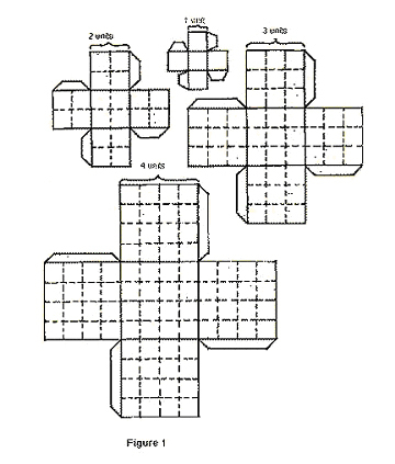
Figure 5. Users explore the architecture of a cell, adjusting size and selecting cell-surface scenarios. (Used by permission of McGraw-Hill.)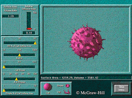
Cell Size Lab - Life Science Resources - Lapeer County, Michigan http://chem.lapeer.org/bio1docs/CellSize.html
In addition to the agar exercise, you may wish to explore the main site ( http://lapeer.org/) which is a community resource for schools and education. Nineteen different educational entities provide Web resources on a countywide level. Ten libraries are also listed as part of this very interesting community site.
How Big Would You Want to Be If You Were a Cell - The SMILE Program http://www.iit.edu/~smile/bi9226.html
The cell diffusion exercise at this Web site is essentially the same as the one above. The main site ( http://www.iit.edu/~smile) is quite extensive. With a project titled “SMILE” (Science and Mathematics Initiative for Learning Enhancement), a team head by Dr. Porter Johnson of the Illinois Institute of Technology of Chicago has assembled over 900 activities and lesson plans involving science and mathematics teaching. This innovative site is worth browsing for teaching ideas, primarily at the 6-12 grade levels.
Figure 6. Sample data available from Cell Size Database.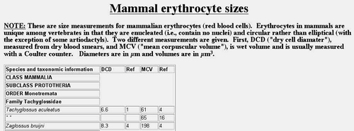
PUTTING THE IDEAS TOGETHER
Textbook publishers such as McGraw-Hill develop interesting support materials for their products. At a commercial Web site titled “Johnson Explorations” the idea of cell size, diffusion, and surface modifications are brought together in an interactive exercise developed for the Raven and Johnson Introductory Biology textbook (ISBN 0-07-303120-8).
Johnson Explorations - Cell Size http://www.mhhe.com/biosci/genbio/biolink/j_explorations/ch02expl.htm
The site opens with a round cell. There are interactive controllers for villi, “dimples”, and cell shape. The surface area to volume ratio as well as diffusion efficiency can be read out. Figure 5 is an example of an adjusted cell. Image courtesy of McGrawHill publishers. (pending) Students can create a number of cell size and cell surface scenarios with the interactive model.
SUGGESTED CELL SIZE LAB
T. Ryan Gregory of the Department of Entomology of London's Natural History Museum has recently assembled what he calls a Cell Size Database. The database contains an extensive collection of vertebrate erythrocyte (red blood cell) sizes arranged by Class, Order, Family, Genus, and species. The database may be found at the following Web site.
Cell Size Database http://www.genomesize.com/cellsize/
Figure 6 represents the information available within the database. Image courtesy of Dr. T. Ryan Gregory, The Natural History Museum, London. Mammalian erythrocytes are generally round with diameters varying from as little as 2.1 μm to as much as 10.8 μm. Other classes of vertebrates have erythrocytes that are generally elliptical with some amphibian species ranging up to 70 by 41 μm in size. In addition to the average size of the cell, the volume is also given. If an instructor is looking for a database that provides information suitable for statistical analysis by students, this would be an excellent place to go. A variety of hypotheses could be developed about Family and Order variation in erythrocyte size. With so many vertebrate species having red cells with diameters in excess of 10 μm, one wishes to see a corresponding database for mean capillary diameters for the same species. A very delicious study could be performed relating capillary size to red cell diameter.
The database contains information about the Mammalian family of organisms known as Camelidae (the camel group). Many people mistakenly think that the erythrocytes of this mammalian family have nuclei, which in the mature form, they don't. Rather, the cells of this mammalian family are elliptical instead of round. Llamas have red blood cells that are 9 by 4.5 μm with a mean volume of 28 μm3. Camel red blood cells are 8.1 by 4.3 μm with a mean volume of 27 μm3. The llama-like vicuna erythrocytes have mean dimensions of 7.1 by 3.9 μm with a volume of 31μ m3.
FUTURE DEVELOPMENTS IN CELL SIZE LABS
The lab exercises referred to in this review suffer from a common shortcoming. None of these exercises explore the relationship between growth and cell division with that of cell size. None of them try to address signaling pathways that lead to the characteristic size of the cell under study. This very rich area of study may be summarized by the earlier cited paper (see reference one in the first section of this article) of Coelho and Leevers (2000) and represented in Figure 7. Image courtesy of Dr. Sally Leevers of the Cancer Research UK London Research Institute.
Besides posing these four possible pathways leading to reproducible cell size, Coelho and Leevers describe the interplay between various cyclins and their regulating kinases. They also explore the rich literature concerning the insulin/P1 3-kinase signaling pathway relationship with cell growth in Drosophila imaginal discs. Day and Lawrence (2000) likewise review the literature dealing with fly wing cell growth (please see reference two in the first section). They also recall the literature about the relationship of ploidy and cell size in salamander. Tetraploid salamanders have cells that are twice the size of diploid salamanders, which leads to interesting results for learning demonstrated in maze tests. Tetraploids have fewer neurons and take longer to learn a route through a maze. The cell size may be different but the body organ size is the same.
Figure 7. Representation of relationship between cell growth and cell division (Coelho, 2000). Reproduced by permission.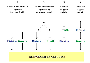
Conlon and Raff (2003) have reported that in rat Schwann cells, cell growth is independent of cell size. They also argue that for Schwann cells the hypothesized cell-size checkpoints operate very differently than has been reported for yeast cells. Hafen and Stocker (2003) ask “Why is an elephant bigger than a mouse?” Their review reminds readers that ribosome biogenesis is an important regulator of multicellular organism cell size.
Web teaching resources dealing with cell size are confined mainly to the physical dimensions of cells. It would be extremely useful to expand the teaching resources to include the signaling events and relationships to the cell cycle. If anyone is aware of such teaching resources please contact me at [email protected]. If you want to comment on the selections or suggest future inclusions, please send a message to the above address.


